Figures & data
Figure 1. Release profile of insulin from chitosan-alginate beads. Beads were incubated in 20 ml release medium (•); artificial gastric fluid, pH 1.2, (◯); artificial intestinal fluid, pH 7.5 on a shaker (100 rpm) at 37°C.
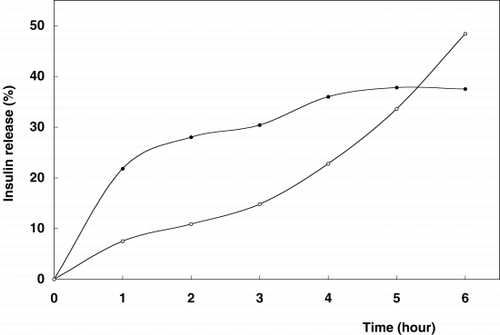
Figure 2. Release profile of insulin from chitosan-alginate-glutaraldehyde beads. Beads were incubated in 20 ml release medium (•); artificial gastric fluid, pH 1.2, (◯); artificial intestinal fluid, pH 7.5 on a shaker (100 rpm) at 37°C.
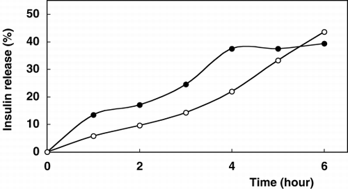
Table 1. In Vitro Insulin Release Towards Enzymatic Degradation Under Gastric Conditions (Artificial Gastric Fluid; pH 1.2 Containing Pepsin at 37°C, 100 rpm)
Table 2. In Vitro Insulin Release Towards Enzymatic Degradation Under Intestinal Conditions (Artificial Intestinal Fluid; TBS pH 7.5 Containing Trypsin at 37°C, 100 rpm)