Figures & data
Figure 1. SEM of the porous scaffolds (a) collagen scaffold without crosslinking, (b) collagen scaffold after cross-linked by formaldehyde, (c) collagen-CS scaffold after cross-linked by EDC.
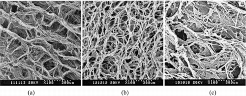
Figure 2. DSC scan of the scaffolds (a) noncross-linking collagen scaffold, (b) formaldehyde cross-linking collagen scaffold, (c) EDC cross-linking collagen-CS scaffold.
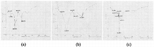
Figure 3. (A) The structure of collagen-CS scaffold soaking in PBS solution. (B) The structure of collagen spongy scaffold soaking in PBS solution. (×250).
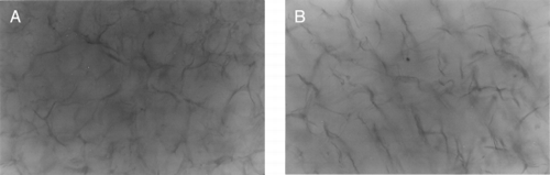
Table 1. Swelling Properties of Collagen and Collagen-CS ScaffoldsFootnotea
Figure 4. Phase contrast micrographs of primary cultured human fetal dermal fibroblasts, cells reached confluence in the (A) second day and (B) fifth day (×250).
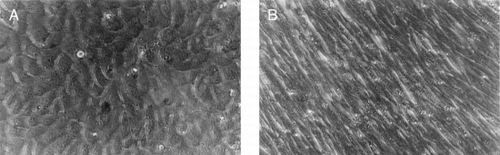
Figure 5. H & E staining of collagen-CS scaffold after fibroblasts implanted for (A) one week, (C) 3 weeks, and (E) 5 weeks. H & E staining of collagen scaffold after fibroblasts implanted for (B) one week, (D) 3 weeks, and (F) 5 weeks. (X250).
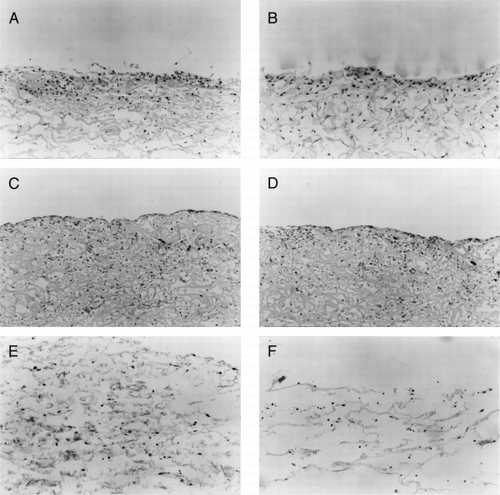
Figure 6. The immunohistological staining of collagen-CS scaffold after fibroblasts cultured for (A) one week, (C) 2 weeks, and (E) 4 weeks. The immunohistological staining of collagen scaffold after fibroblasts cultured for (B) one week, (D) 2 weeks, and (F) 4 weeks. (X250).
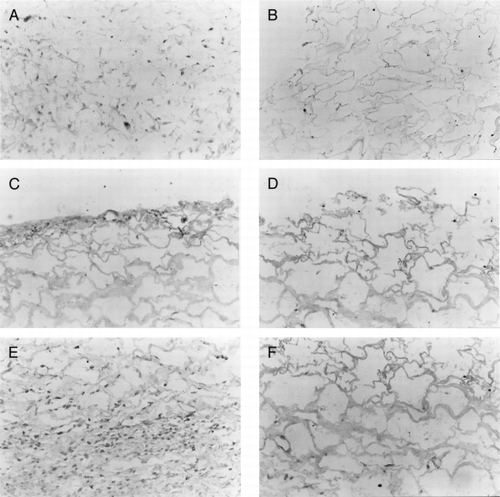
Table 2. The Stains of the FN and Type I Collagen of the Two ScaffoldsFootnotea
Table 3. The GAG Content in Two Scaffolds μg/well
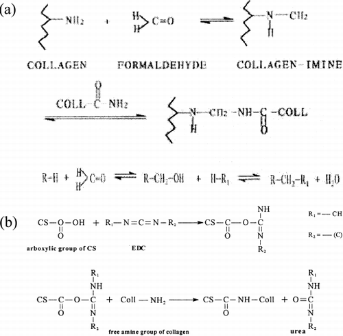
Figure 7. (a) Reaction mechanism of formaldehyde crosslinking the collagen. (b) Reaction mechanism of EDC crosslinking the collagen and EDC.