Figures & data
Table 2. Enzymes and Proteins Enclosed in Artificial Cells
Figure 29. Urease activity, measured as rate of rise of pH of urea-buffer medium. No pH change in the presence of supernatant from a 50% suspension of artificial cells stored for 12 hours. The artificial cells are homogenized and separated into the membrane fraction and homogenate fraction. Nearly all the enzyme activity is located in the membrane-free homegenate.
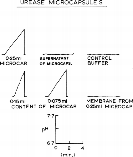
Figure 30. Stability of urease enclosed in nylon membrane artificial cells, stored in phosphate buffer at pH 6.7. Continuous lines represent activity after storage at 0°C (upper line) and at 37°C (lower line) for enzyme encapsulated with hemolysate. Discontinuous line represents activity after storage at 37°C for enzyme encapsulated without hemolysate. (From Chang, 1965.)
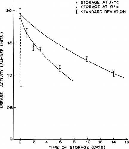
Figure 31. Schematic representation of the action of intraperitonally injected artificial cells loaded with urease. (From Chang, 1965.)
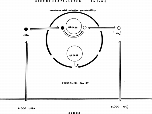
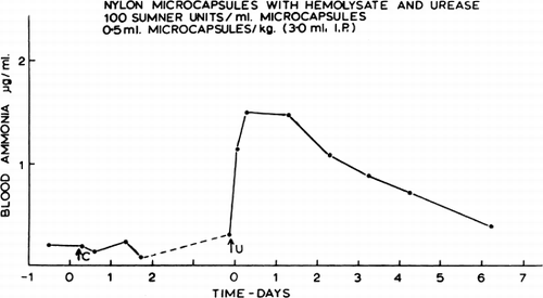
Figure 32. Effect of urease-loaded artificial cells on blood ammonia in dogs anesthetized with Nembutal; the graph summarizes data from 3 experiments (mean±SD). C=injection of control artificial cells (no urease, 0.25 ml/kg). U=injection of urease-loaded artificial cells (0.25 ml and 100 Sumner units/kg). (From Chang, 1965.)
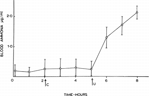
Figure 33. Effect of artificial cells on blood ammonia in an unanesthetized dog. C=control artificial cells (0.5 ml/kg). U=urease-loaded artificial cells (0.5 ml and 50 Sumner units/kg). In this test, the urease preparation (NBC) was stabilized by the addition of hemolysate. (From Chang, 1965.)