Figures & data
Figure 1. Plasma clearance and half‐life (T1/2) of total Hb raffimer (THb) administered to dogs subjected to a single topload infusion of 18 mL/kg. Total hemoglobin values are shown by the filled circles (•). Mean plasma methemoglobin (MetHb) levels are given by the (*) symbol. All values are presented as mean ± SD.
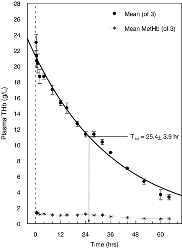
Figure 2. Plasma clearance and half‐lives (T1/2) of the different hemoglobin fractions that make up Hb raffimer as determined by size exclusion chromatography under dissociating conditions. The disappearance of the 64 kDa fraction (tetramer) is shown in the top panel, the 128 kDa fraction in middle panel, and the fraction composed of all molecular weight species > 128 kDa in the bottom panel. All values are shown as mean ± SD for all animals.
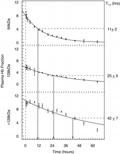
Figure 3. The relative percentage of the different hemoglobin fractions that make up Hb raffimer in the plasma over time. Grey bars indicate the 32 kDa fraction, hatched bars indicate the 64 kDa fraction and open bars indicate > 64 kDa fraction.
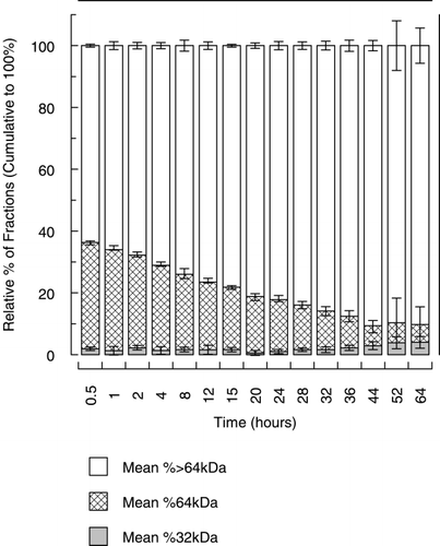
Figure 4. Reverse phase chromatogram showing absorbance at 220 nM. Chromatograms presented are from a control beagle dog, human hemoglobin A0, the 32 kDa fraction of Hb raffimer and a 32 kDa fraction collected from a dog 52 hours after treatment with Hb raffimer. For this sample the scale is expanded 2 ×.
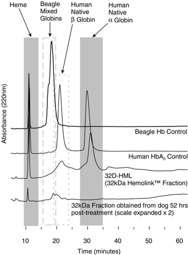
Figure 5. Plasma MetHb levels (•), expressed as a percentage of the total plasma hemoglobin concentration (top panel) and also presented as the plasma mass concentration in g/L (bottom panel). Levels are not reported beyond 32 hrs as hemoglobin concentration has fallen below 10 g/L and measurement of MetHb is not accurate at such low levels of total plasma hemoglobin. For comparison, total plasma hemoglobin concentration (○), expressed in g/L is also shown on the right Y‐axis. All values presented are given as mean ± SD.
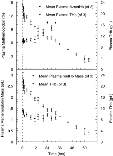
Figure 6. Whole blood methemoglobin levels (•), expressed as a percentage of the total whole blood hemoglobin concentration (top panel) and also presented as the whole blood mass concentration in g/L (bottom panel). For comparison, total whole blood hemoglobin concentration (○), expressed in g/L is also shown on the right Y‐axis. All values presented are given as mean ± SD.
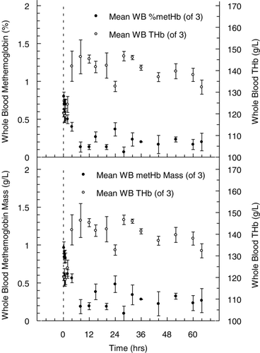
Table 1. Mean urine hemoglobin concentrations (g/L)