Figures & data
Figure 1. The surface of the hollow fiber of polypropylene K600®PP, Accurel, Akzo original (× 1000).
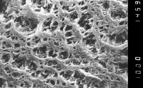
Figure 2. The surface of the hollow fiber of polypropylene K600®PP, Accurel, Akzo modified by siliconization (× 1000).
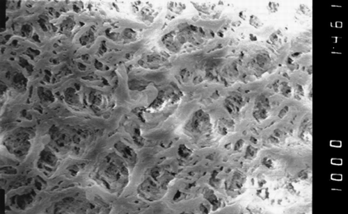
Figure 3. The dialyser circuit.
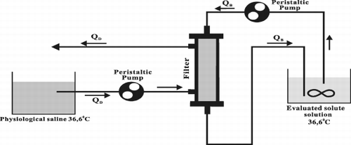
Table 1. Diffusive Permeability Values for Polypropylene K600 PP, Accurel Original or Silikonized HF for Creatinine, Bovine Albumin, Human Iodinated Albumin, IgG and Myosin Measured for Single HF and for HF in Module. The Values of the Rate of HF Diffusion Coefficient to Water Coefficient for Examined Solutes (Dmem/DH2O)
Figure 4. The product of diffusion coefficient and particle radius in the function of particle radius in water and in permiselective membrane for creatinine, IgG, myosine.
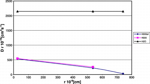
Figure 5. Relationship between particle radius and the product of particle radius and diffusion coefficient, calculated and experimental.
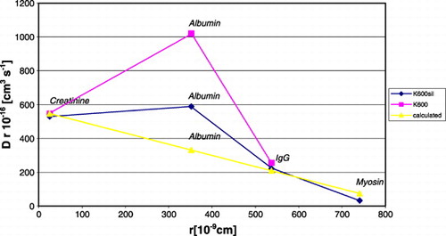
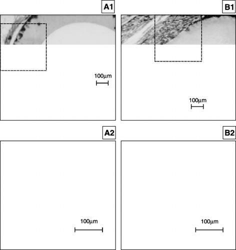
Figure 6. A1, A2. The view of the K600 siliconized HF surface after 105 days implantation into mouse. The external surface of HF was surrounded with the thin layer of fibroblasts (thickness about 30µm), there was calcium salt intrusion in the membrane wall. There was a slit between the fibroblasts layer and the external HF surface. The implant appeared to be accepted (× 70). B1, B2. The view of the K600 original HF surface after 105 days implantation into mouse. There was foreign body reaction on the surface of the HF. The presence of macrophages, fibroblasts and lymphocytes was observed (× 70).