Figures & data
Figure 1. LS signals of BSA at different concentration. c1 to c5 is 0.4748, 0.5953, 0.9427, 1.196, 1.909 mg/mL, respectively. (View this art in color at www.dekker.com.)
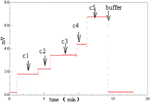
Figure 2. Debey plot of MALS signals to determine molecular weight of BSA at different sample concentrations. MALS signals at each concentration of BSA in were replotted by Debey plots with Astra software.
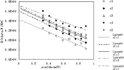
Figure 3. SEC chromatogram of BSA. Ten microliters of 4 mg/mL BSA was injected onto a G4000SW column (Tossohaas) with PBS as eluant at 0.5 mL/min flow rate. The solid line is the RI signal and the dashed line is the LS signal. (View this art in color at www.dekker.com.)
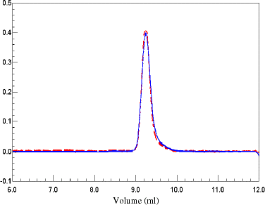
Figure 4. RI signals and MW of BSA. Molecular weights were calculated by Debey plot. The molecular weights were in the range from 64–67 kDa. (View this art in color at www.dekker.com.)
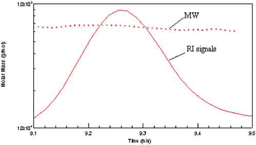
Figure 5. Molecular weight as a function of concentration of bHb using and methods. ▴ represents the molecular weights calculated from microbatch MALS data; ♦ represents the molecular weights calculated from the first half of peak of online SEC-LS/RI data; and −represents the molecular weights calculated from the second half of peak of online SEC-LS/RI data.
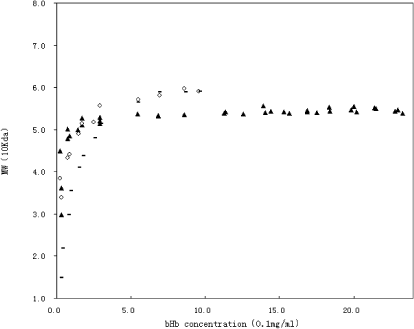
Figure 6. SEC chromatogram of bHb. One hundred microliters of 4 mg/mL bHb was injected onto a G4000SW column (Tossohaas) with PBS as eluant at 0.5 mL/min flow rate. The solid line is the RI signal and the dashed line is the LS signal. (View this art in color at www.dekker.com.)
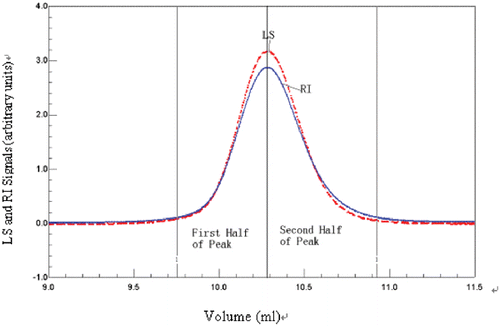
Figure 7. Light-scattering signals of bHb at different pH. BHb concentration is 1.85 mg/mL in different buffer. The buffer is 0.15 M NaCl and 0.05 M phosphate, and the pH ranges from 2.85 to 11.41 (2.85, 5.30, 5.52, 6.17, 6.96, 7.99, 8.79, 9.96, 11.71, respectively). (View this art in color at www.dekker.com.)
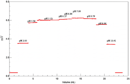
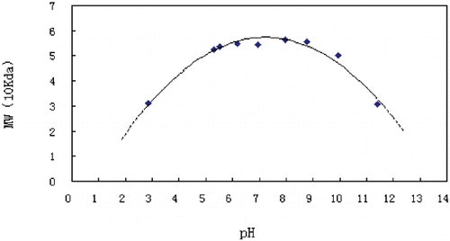
Figure 8. Effect of pH on the molecular weight of bHb. Molecular weights at 1.85 mg/mL of bHb, calculated by the Debey plot, is plotted against pH of the buffer, 0.15 M NaCl and 0.05 M phosphate.