Figures & data
Figure 1. (a) A typical elution pattern from the TSK G3000SWXL column for the purified stroma-free hemoglobin solution; (b) Electrophoresis in SDS-page of the purified stroma-free hemoglobin solution.
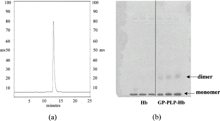
Figure 2. (a) A typical HPLC molecular weight profile following 4 h after initiation of polymerization of hemoglobin by genipin; (b) A typical HPLC molecular weight profile following 30 min after initiation of polymerization of hemoglobin by glutaraldehyde at the same reaction conditions.
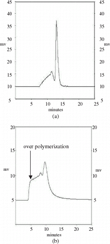
Table 1. The durations taken (h) for various reaction conditions (temperature, hemoglobin concentration, and genipin-to-hemoglobin molar ratio) to achieve the maximum degree of hemoglobin polymerization by genipin (˜40%)
Table 2. Percentages of methemoglobin produced at various reaction conditions investigated (temperature, hemoglobin concentration, and genipin-to-hemoglobin molar ratio) after the maximum degree of hemoglobin polymerization by genipin was achieved
Figure 3. Effects of using glycine at various concentrations (in glycine-to-hemoglobin molar ratio) on the termination of hemoglobin polymerization by genipin.
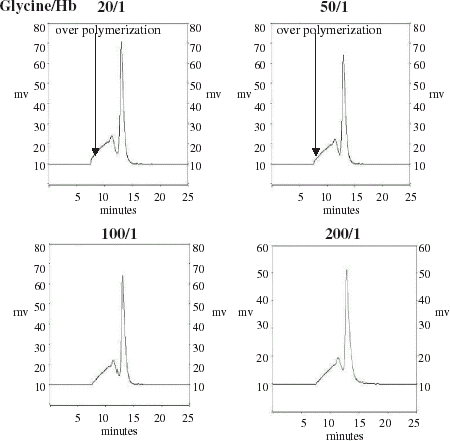
Figure 4. Results of removal of the unpolymerized hemoglobin (PLP-Hb) from the genipin-polymerized hemoglobin (GP-PLP-Hb) carried out by an ion-exchange column (b) or a gel-filtration column (c).
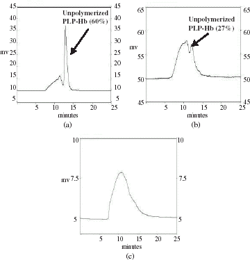
Figure 5. Particle-size distribution curves for the unmodified Hb and GP-PLP-Hb solutions determined by a light-scattering method. (View this art in color at www.dekker.com.)
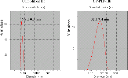
Table 3. Characteristics of the unmodified Hb and GP-PLP-Hb solutions used for exchange transfusions in the rat
Table 4. Results of the first group of experiments in examining the survival of the rats at an approximately 50% blood-volume-exchange transfusion with the unmodified Hb, PBS, or GP-PLP-Hb solution
Figure 6. Disappearance of plasma hemoglobin for the animals treated with the unmodified Hb or GP-PLP-Hb solution.
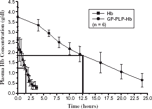
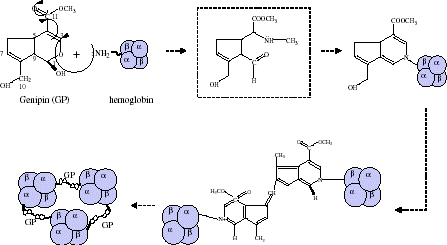
Figure 7. Presumable mechanism of hemoglobin polymerization by genipin. (View this art in color at www.dekker.com.)