Figures & data
Figure 1. Degradation of the B epitope as a function of enzyme concentration. A purified preparation of R. gnavus α-galactosidase was serially diluted into PBS, pH 7.0. Enzyme treatment of B membrane coated microplates and ELISA assay were performed as previously described. Error bars indicate range of ΔOD415. All data points are the product of three independent duplicate determinations.
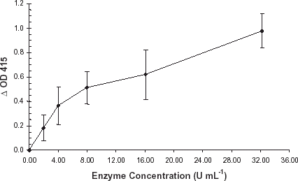
Figure 2. H antigen expression. A purified preparation of enzyme was diluted with PBS, pH 7.0, and applied to a B membrane coated plate. Negative controls consisted of PBS, pH 7.0, incubated under the identical conditions. Enzyme treated wells and negative controls were developed per normal procedure. Primary antibody consisted of monoclonal anti-B (1:100) or monoclonal anti-H (1:10). Error bars indicate range of OD415. All data points are the mean of two independent duplicate determinations.
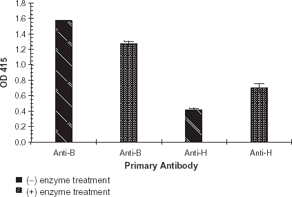
Figure 3. Diluted red cell preservative study: Adsol, CPDA-1, and CPD. Red cell preservative solutions Adsol, CPDA-1, and CPD were diluted 1:10 with PBS, pH 7.0. A purified preparation of enzyme was diluted with each of these solutions. The enzyme treatment and ELISA plates were developed per normal procedure. Bars indicate change in OD415. Error bars indicate range of ΔOD415. Results are the mean of two independent duplicate determinations.
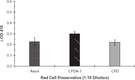
Figure 4. Red cell preservative pH study. Red cell preservative CPD, pH 6.25, 6.50, 6.75 and 7.0 was prepared. A purified preparation of enzyme was diluted with each of these solutions. The dilutions were applied to B membrane coated ELISA plates and developed per normal procedure. Bars indicate change in OD415. Error bars indicate range of ΔOD415. Results are the mean of two independent duplicate determinations.
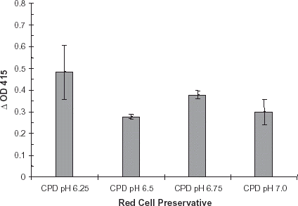
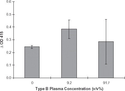
Figure 5. Effect of type B plasma: Red cell preservative solution CPD, pH 7.0, was diluted 1:10 with PBS, pH 7.0. This solution was used to dilute type B plasma to the following concentrations: 0%, 9.2% and 91.7% plasma (see Methods). A purified preparation of enzyme was diluted with each of the plasma solutions. Negative controls consisted of identical plasma dilutions minus enzyme. The ELISA assays were performed per routine. Error bars indicate range of ΔOD415. Results are the mean of two independent duplicate determinations.