Figures & data
Figure 1. Set-up of in vitro measurements to determine absorber accuracy and volume error of the CO2SMO pneumotachograph using a syringe. By modifying the PFC infusion speed the PFC content in the breathing gas could be altered and its influence on volume measurement be analyzed.
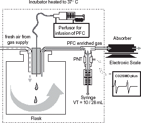
Figure 2. Adsorption curve of the absorber over time while delivering 5 mL PFC with a speed of 15 mL h−1. Depicted are the weight of the absorber (top) and the weight gain (bottom) over time. During PFC infusion the weight of the absorber increased linearly until a plateau is reached.
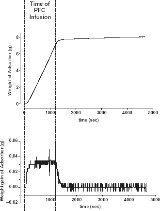
Figure 3. Measurement error of the evaporated PFC volume calculated from the weight gain of the absorber.
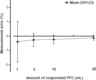
Figure 4. Measurement error of the PFC flow rate calculated by differentiation of the weight gain curve.
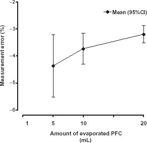
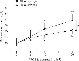
Figure 5. (p < 0.05) marks statistically significant differences between both tidal volumes.