Figures & data
Figure 1 Effect of HbV on platelet surface activation markers. (A) PAC-1 binding to platelets and (B) CD62P expression on platelets. Whole blood was incubated with HbV at concentrations of 0% (square), 20% (triangle), or 40% (circle). Whole blood was then stimulated with or without various concentrations of ADP, as described in the Materials and Methods section. Values are the means±SE of 4 experiments. *p < 0.05, **p < 0.01, compared with control (0% HbV).
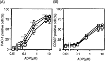
Figure 2 Effect of HbV on ADP-induced platelet mediator release. ADP-induced release of (A) TXB2, (B) RANTES, and (C) β-TG from human platelets. PRP was incubated with concentrations of 0%, 20%, or 40% HbV and then stimulated with (hatched columns) or without (open columns) ADP, as described in the Materials and Methods section. Values are the means±SE of 5 (A) and 6 (B, C) experiments using blood from different donors. *p < 0.05, compared with control (0% HbV).
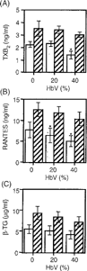
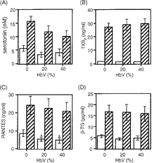
Figure 3 Effect of HbV on collagen-induced platelet mediator release. Collagen-induced release of (A) serotonin, (B) TXB2, (C) RANTES, and (D) β-TG from platelets. PRP was incubated with concentrations of 0%, 20%, or 40% HbV and then stimulated with (hatched columns) or without (open columns) collagen, as described in the Materials and Methods section. Values are the means±SE of 5 experiments using blood from different donors. *p < 0.05, compared with control (0% HbV).