Figures & data
Figure 1 Graph to demonstrate concentration of free iron in plasma derived from blood samples taken from rats at various times after bolus intravascular injection of 5 ml DBBF-Hb (50 mg) or isotonic saline. Error bars indicate SEM. After 15 min. there was significantly more iron in the samples as a result of the DBBF-Hb injection compared to control values (88.07 ± 3.75 (SEM) µg/dL vs. 61.36 ± 2.00 (SEM) µg/dL, p < 0.05.
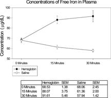
Figure 2 Average number of intestinal mucosal degranulated mast cells per villus for different treatment. The treatments consisted of a 0.5 ml bolus injection of the first substance, followed 2 minutes later by a 5 ml bolus injection of the second substance. All experiments were terminated 30 minutes after the second injection. In all cases, the concentrations of DBBF-Hb, iron dextran and PS-ODN were 10 mg/ml, 0.9 µg/ml and 1 mg in 0.5 ml, respectively; *Significantly larger than saline control group, p < 0.05; #Significantly smaller than saline/DBBF-Hb group at same time point, p < 0.05.
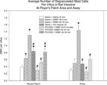
Figure 3 Average number of intestinal mucosal secreting goblet cells per villus for different treatments as described for Figure 2. In all cases, the concentrations of DBBF-Hb, iron dextran and PS-ODN were 10 mg/ml, 0.9 µg/ml and 1 mg in 0.5 ml; *Significantly larger than saline control group, p < 0.05; #Significantly different from saline/DBBF-Hb group at same time point, p < 0.05.
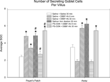
Figure 4 Electron microscopy of intestinal mucosa to assess epithelial damage. Typical electron micrograph of transverse section through intestinal villus (away from Peyers' patch) from rat injected with a 5 ml bolus of HBS-2% BSA (Hepes buffered saline containing 2% bovine serum albumin). The epithelium (E) is mostly intact and an intact mast cell (MC) is visible. Capillary cross-sections (C) can also be seen. Villi close to Peyers' patches were similar in appearance; Scale bar: 5 microns.
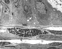
Figure 5 Electron microscopy of intestinal mucosa to assess epithelial damage. Typical electron micrograph of transverse section through intestinal villus (Peyer's patch area) 30 minutes after injection of rat with a 5 ml bolus of iron dextran (0.9 µg/ml). The epithelial cells (E) are separating from each other and from the basement membrane (arrows) and show extensive cytoplasmic protrusions. The interstitium, (I) is edematous. C: capillary containing two red blood cells; Scale bar: 5 microns.
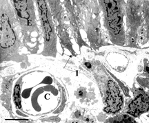
Figure 6 Electron microscopy of intestinal mucosa to assess epithelial damage. Typical electron micrograph of transverse section through intestinal villus (away from Peyer's patch) from rat that had been injected with 1 mg PS-ODN in 1 ml, followed 2 minutes later by 50 mg DBBF-Hb in 5 ml. The experiment was terminated eight minutes after the DBBF-Hb injection. Similar to the results of injection with iron dextran (Figure 5) and with DBBF-Hb alone (not shown), the epithelial cells (E), with extensive cytoplasmic protrusions, are separating from each other and from the basement membrane (arrows). Near Peyers' patches, the areas of epithelial disruption were even more extensive than with DBBF-Hb alone. C: capillary containing two red blood cells. Scale bar: 5 microns.
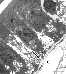
Figure 7 Electron microscopy of intestinal mucosa to assess epithelial damage. Typical electron micrograph of transverse section through intestinal villus (away from Peyer's patch) from rat that had been injected with 0.5 ml HBS-2% BSA, followed 2 minutes later by 50 mg DBBF-Hb. The experiment was terminated 30 minutes after the DBBF-Hb injection. The epithelial cells (E) are much closer together than after 8 minutes perfusion with DBBF-Hb, and the cytoplasmic protuberances are much less evident, indicating some repair of the epithelial lining. Villi near Peyer's patches were even more intact. Scale bar: 5 microns.
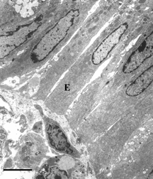
Figure 8 Electron microscopy of intestinal mucosa to assess epithelial damage. Typical electron micrograph of transverse section through intestinal villus (Peyers' patch area) from rat that had been injected with 1 mg PS-ODN in 0.5 ml, followed 2 minutes later by 50 mg DBBF-Hb in 5 ml. The experiment was terminated 30 minutes after the DBBF-Hb injection. Unlike the villi from animals injected with HBS-2% BSA for 2 minutes followed by DBBF-Hb for 30 minutes (Figure 7), the villous epithelial cells (E) are separated from each other and show extensive cytoplasmic protuberances. A degranulated mast cell (DMC) is visible. Thus pretreatment with PS-ODN appears to inhibit epithelial repair of villi adjacent to Peyer's patches. Scale bar: 5 microns.
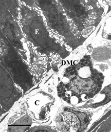
Figure 9 The average Epithelial Integrity indices for each group. The scale ranges from 1 to 3; a score of 1 means the cells are intact, 2 means the cells show some separation from each other, and 3 means there is some separation of cells from the basement membrane; *Significantly larger than corresponding saline control group, p < 0.05; #Significantly different from saline/DBBF-Hb group at same time point, p < 0.05.
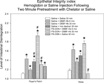
Figure 10 In vivo relative estimation of reactive oxygen species in intestinal mucosa. Histogram to show fluorescence intensity of dihydrorhodamine 123 in the epithelium (upper panel) and lamina propria (lower panel) of intestinal villi after injection of DBBF-Hb (5 ml bolus at 10 mg/ml), following pre-treatment with saline (0.5 ml HBS-2% BSA for 2 minutes) or chelator (0.5 ml PS-ODN (1 mg) for 2 minutes). On the abscissa is plotted the time after injection of DBBF-Hb in minutes. Asterisks signify value is significantly greater for chelator pre-treatment than for saline pre-treatment.
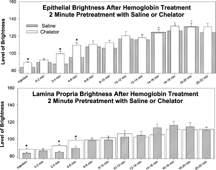
Table 1. Percentages of different oxidation states of DBBF-Hb under various conditions