Figures & data
Figure 1 Limited enzyme incision confirmation map of report vector pGFAP-EGFP-N3. 1 lane: λDNA/Hind III maker, 2 lane: Xho I and Sal I digested fragments, 3 lane: XbalI digested fragments; 4 lane: EcoR I digested fragments. There were three Xbal I limited enzyme incise sites in reconstructed vector and one site was methylated down EGFP, which cannot be digested.
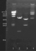
Figure 2 Immunofluoresence staining of induced MSCs (100 ×). A: GFAP as primary antibody; B: S100 as primary antibody; (Olympus XI 70, Japan).
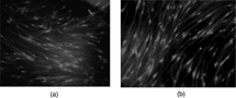
Figure 3 RT-PCR electrophoresis results. 1 lane: DNA maler; 2 and 4 lanes: GFAP; 3 and 5 lanes: S100. The deviced GFAP sequence to be amplified was about 270 bp, and the S100 sequence was about 300 bp.
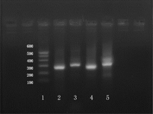
Figure 4 Flow cytometry analysis of EGFP expression. Upper was the control group; lower was the EGFP expression rate.
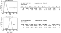
Figure 5 The EGFP-expressing MSCs (green) mainly accumulated around the injured nerve fiber stem (100 ×).
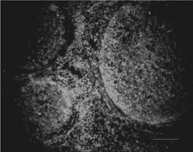
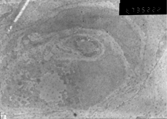
Figure 6 Immuno-electron microscope morphyological observation of MSCs (6700 ×) (JEOL100CX-II, Japan).