Figures & data
Figure 1. PA chest radiograph at presentation (left) and then 2 years later (right). Both radiographs demonstrate typical changes associated with emphysema, including hyperinflation, flattening of the diaphragm, and a paucity of pulmonary vascular markings peripherally. Note the rapid development of large bullous disease on the right.
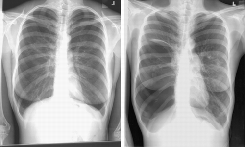
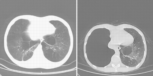
Figure 2. CT scan of the thorax, at presentation (left) and 2 years later (right), again demonstrating the rapid development of a large bulla in the right hemithorax with shift of the heart to the left.