Figures & data
Figure 1 Block copolymer nanoassemblies reported for tumor targeted drug and gene delivery.
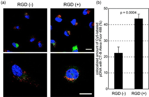
Figure 2 (a) Chemical structure of PEG-b-PAsp(DET) copolymer bearing an ethylenediamine unit at the side chain, which can form stable polyplexes with pH-sensitive protonation properties. (b) The protonation change of the ethylenediamine in the PAsp(DET) backbone of the polyplexes leads to the membrane destabilization at the acidic pH of late endosomal compartments facilitating the efficient translocation of the polyplexes to cytoplasm.
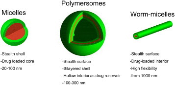
Figure 3 PIC micelles sensitive to reductive conditions. Change in the relative scattering light intensity of disulfide-crosslinked PIC complex micelles after the addition of dithiothreitol (DTT) (○, 0.5 mM; ▵, 1.0 mM; □, 2.0 mM) in 10 mM PBS at pH 7.4. (Reprinted with permission from [Citation30] © 1999 American Chemical Society.)
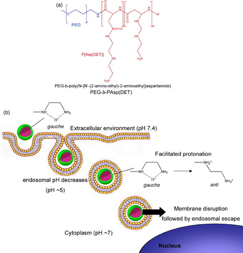
Figure 4 Photosensitive block copolymer for nanoassemblies. Chemical structure of PEG-b-poly([7-(diethylamino)coumarin-4-yl]methyl methacrylate) and photolysis reaction after UV or NIR irradiation.
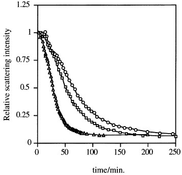
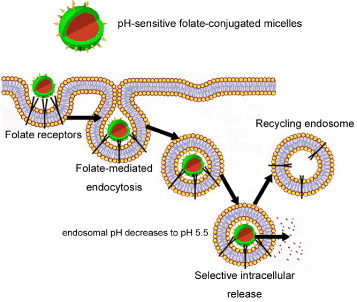
Figure 5 Folate targeted nanoassemblies. Enhanced cellular uptake by folate-mediated endocytosis of folate-conjugated nanoassemblies. The plasma membrane envelops the nanoassembly/folate receptor complex to form an endosome. As the lumen of the maturing endosome acidifies up to pH 5.5, the receptor changes the conformation and releases the conjugate.
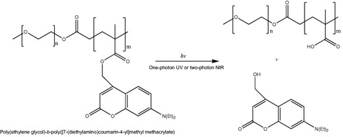
Figure 6 Intracellular distribution of cRGD-conjugated disulfide-crosslinked PIC micelles. Polyplex micelles loading Cy5-labeled pDNA (red) and CT-B Alexa Fluor 488 conjugate, a marker for the lipid rafts and the caveosomes (green), were incubated with HeLa cells for 1 h, washed and reincubated for 11 h. The cell nuclei were stained with Hoechst 33342 (blue). (a) CLSM images of RGD (−) micelles (left) and RGD (+) micelles (right). The scale bars represent 20 μm. (b) Quantification of Cy5-labeled pDNA colocalized with CT-B in the inner-cytoplasm. Error bars represent SEM (n=10). (Reprinted with permission from [Citation72] ©2008 The American Society for Pharmacology and Experimental Therapeutics.)