Figures & data
Scheme 1 Preparation schemes of pliable biodegradable tubes with microchannels. Four-armed P(CL-co-DLLA)s are synthesized by ring-opening copolymerization of CL and DLLA from terminal hydroxyl groups of pentaerythritol. Acryloyl chloride is then reacted with the ends of the chains. The end-functionalized P(CL-co-DLLA) is crosslinked in a cylindrical mold in the presence of longitudinally oriented silica fibers as the templates, which are later dissolved with HF. SEM images of the silica fibers (a) and the crosslinked P(CL-co-DLLA) before (b) and after etching (c).
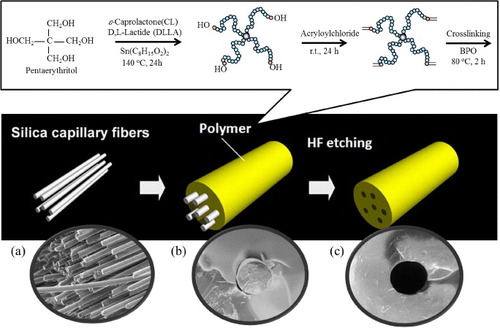
Figure 1 (a) Schematic of crystal–amorphous transition of semicrystalline PCL. (b) Effect of DLLA content in crosslinked P(CL-co-DLLA) on the melting temperature (Tm) determined by DSC.
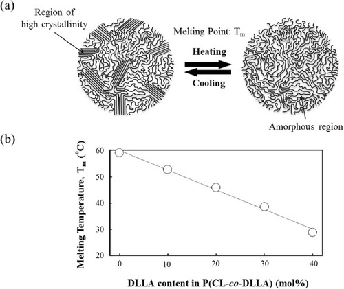
Table S1. Properties of four-armed P(CL-co-DLLA).
Figure 2 Temperature dependences of Young's modulus for crosslinked PLLA and P(CL-co-DLLA). Solid circles: PLLA, open squares: 80/20, open triangles: 70/30, open circles: 60/40 PCL/DLLA.

Figure 3 (a) Representative SEM images of microchannel structures produced in P(CL-co-DLLA) (70/30) scaffolds using silica templates with diameters 8, 150, 350 and 660 μm. (b) Relationship between diameters of silica templates and channels created in the scaffolds.
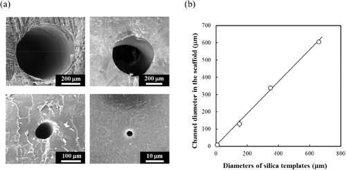
Figure 4 Optical (a) and electron (b,c) micrographs of P(CL-co-DLLA) (70/30) (left) and PLLA (right) tubes with inner diameters of 350 μm. (a) The samples were bent to 90° at 37 °C. Cross-sectional images of the tubes (b) and PDMS replica (c) at the bending point.
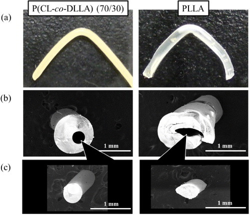
Figure 5 The effects of channel diameter on Young's modulus and stress at break of P(CL-co-DLLA) (70/30) at 37 °C. All samples possess the same 38% porosity which was adjusted by the number of templates: for 150, 350 and 660 μm diameters, 39, 7 and 2 channels were created in the scaffold, respectively.
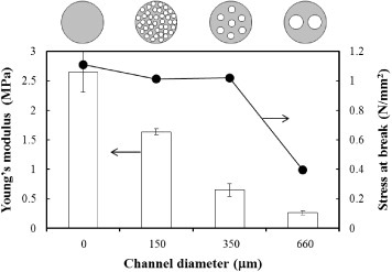
Figure 6 (a) Schematic of experimental setup for cell migration assay. Transparent PDMS microchannels enable the microscopic observation of Schwann cells migration in the channel. We measured the migration distances of Schwann cells on the poly-L-lysine-coated TCPS, which was covered with PDMS channels of various widths (b) and heights (c).
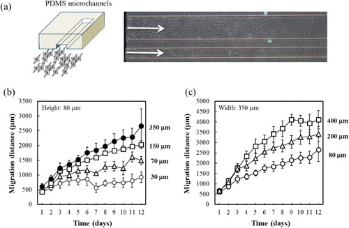
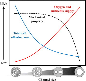
Figure 7 Schematic of the overall trends of the channel size effects on mechanical properties (dashed black line), total surface area available for cell adhesion (solid blue line), and individual channel volume to supply oxygen and nutrients (solid red line).