Figures & data
Figure 1. Chemical structure of AGMA1 repeating unit and RGD motif.
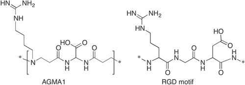
Figure 2. Representative bright field microscopy images of primary rat microglia grown on PLO, PLL and AGMA1. Scale bar = 25 μm.
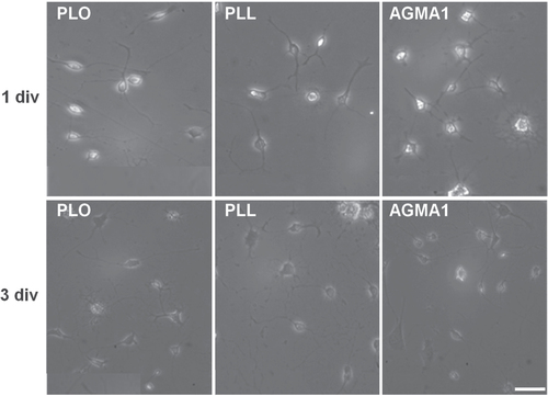
Figure 3. Representative confocal microscopy pictures of primary mixed coculture neurons-astrocytes grown on AGMA1 (A) and PLL (B). Scale bar = 25 μm.
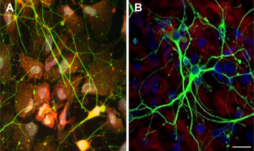
Figure 4. Representative bright field microscopy pictures at different times of primary rat hippocampal neurons grown on AGMA1 and PLL. Scale bar = 25 μm.
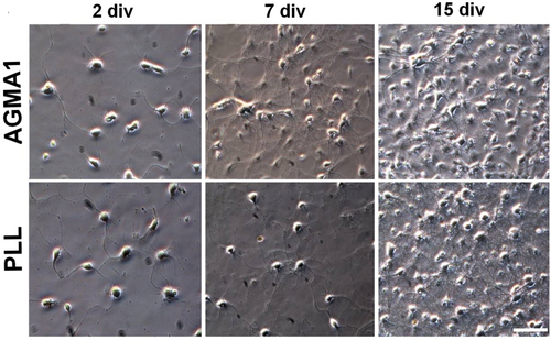
Figure 5. Representative confocal microscopy pictures of primary hippocampal neurons stained with early marker MAP-2 at 2, 7 and 10 div and with both MAP-2 and synaptic vesicle associated marker VAMP-2 at 10 div. Differential interference contrast (DIC) images at 2 and 7 div. Scale bar = 25 μm.
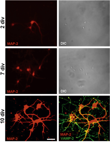
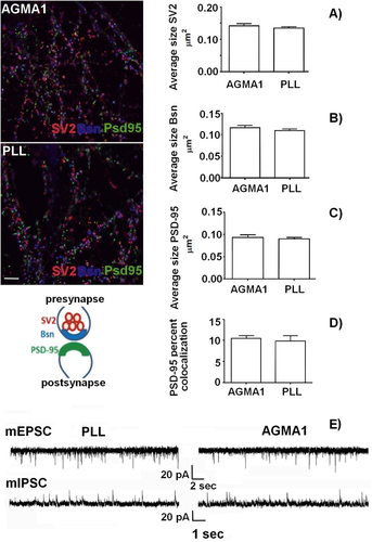
Figure 6. Immunofluorescence staining of 16 div primary hippocampal neurons plated on AGMA1 and PLL with synaptic markers SV2 (red), Bassoon (Bsn, blue) and PSD-95 (green). Quantitative analysis: average size of SV2 (A), Bassoon (B) and PSD-95 (C) puncta. (D) Percentage of area of PSD-95 puncta colocalized with SV2 and Bassoon vs total area of PSD-95 puncta. No significant difference in all parameters is detectable between cultures plated on AGMA1 or PLL. The cartoon illustrates the relative localization of the three markers at the synapse. Scale bar = 25 μm. (E) Representative traces of mEPSCs and mIPSCs recorded from 16 div hippocampal neurons plated on PLL substrate or AGMA1 showing the occurrence of miniature events in both experimental conditions, indicating that neurons cultured on AGMA1 are characterized by spontaneous activity comparable to that of neurons grown on PLL coated medium.