Figures & data
Table 1. Composition of 3D nanocomposite scaffolds.
Figure 1. Quantitative flow cytometry analysis of typical cell surface markers (red color) from mesenchymal stem cell (CD44, CD90, and CD105) (a)–(c) and hematopoietic stem cell (CD45) (d).
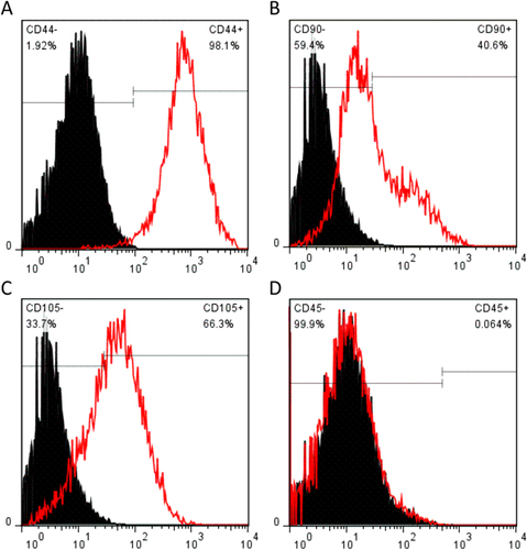
Figure 2. Multilineage differentiation of SMSCs. (a) After incubation in chondrogenic medium SMSCs were positively stained for collagen II protein (immunostaining). (b) After incubation in adipogenic medium SMSCs were positively stained for adipose drops (Oil Red O). (c) After incubation in osteogenic medium SMSCs were positively stained for calcium matrix (Alizarin Red).
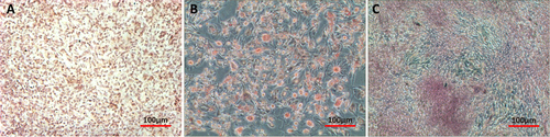
Figure 3. SEM images of gelatin nanoparticles.
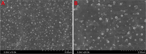
Figure 4. 3D printed nanocomposite scaffolds. (a) NP/SF, (b) NP/SF–Gel, (c) NP/Gel-1, (d) NP/Gel-2.
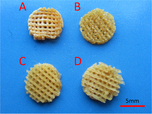
Figure 5. SEM images of 3D printed nanocomposite scaffolds. (a) NP/SF, (b) NP/SF–Gel, (c) NP/Gel-1, (d) NP/Gel-2.
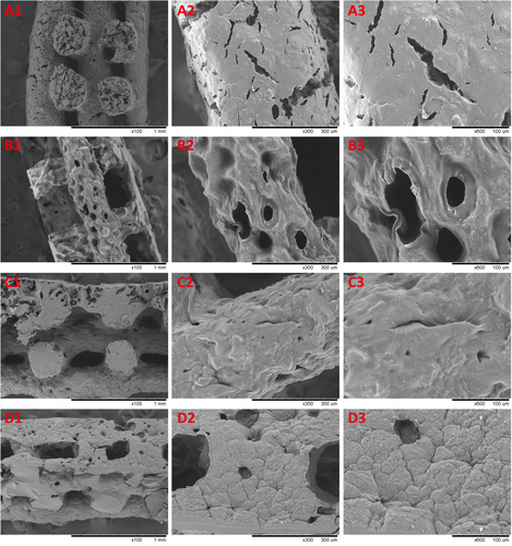
Figure 6. The proliferation of SMSCs cocultured with 3D printed nanocomposite scaffolds in the CCK-8 assay: the absorbance of these medium with CCK-8 was read at 450 nm.
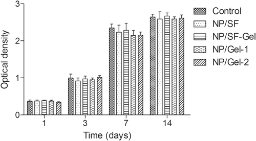
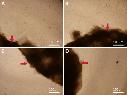
Figure 7. Microscopy images of SMSCs cocultured with 3D printed nanocomposite scaffolds shown by red arrows (a) NP/SF, (b) NP/SF–Gel, (c) NP/Gel-1, (d) NP/Gel-2.