Figures & data
Figure 1 Molecular structures of the glycopolymers with trehalose and sugar alcohols.
Molecular weights of the glycopolymers.a
Figure 2 Effect of trehalose (1) and polyvalent trehalose (2) on Aβ (1–42) aggregation. Control (∘), 1 (•) and 2 (□).
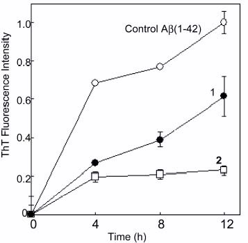
Figure 3 Effect of glycopolymer (3, 4, 5 and 6) on Aβ(1–42) aggregation. Control (∘), 3 (▴), 4 (▪), 5 (X), 6 (✦), and 7 (◊).
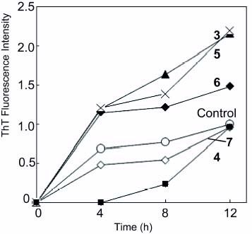
Figure 4 AFM of Aβ(1–42) (a) without glycopolymer, (b) in the presence of 2 and (c) in the presence of 3. Control sample of glycopolymer (2) without Aβ(1–42) (d).
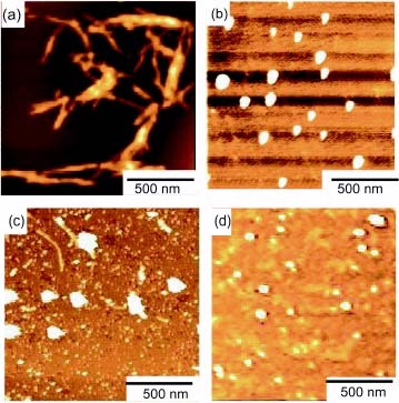
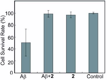
Figure 5 Neutralization of Aβ(1–42) with a glycopolymer (2). The survival rate of HeLa cells against Aβ(1–42) were monitored in the presence of the glycopolymer. Results were the average of at least three samples.
The diameters of glycopolymers in aqueous solution.
Figure 6 Correlation between diameter of glycopolymer and aggregation properties of Aβ(1–42). The diameter of the glycopolymer was plotted against ThT fluorescence intensity after 8 h incubation.
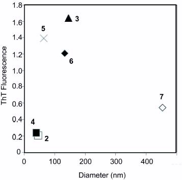
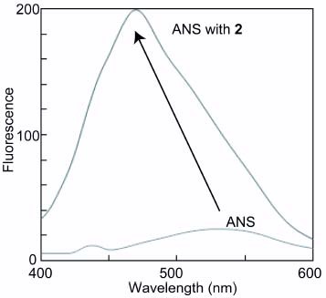
Figure 7 Fluorescence spectra of ANS in 2 (100 μM) with excitation wavelength at 345 nm.