951
Views
30
CrossRef citations to date
0
Altmetric
ORIGINAL ARTICLE

Localization of acid phosphatase activities in the roots of white lupin plants grown under phosphorus-deficient conditions

, , , , &
Pages 95-102 | Received 29 Jun 2007, Accepted 28 Aug 2007, Published online: 21 Dec 2010

Figures & data

Figure 1  Activity staining of APase separated by isoelectric focusing. (A) Profile of APase isoforms in crude protein extracted from roots of hydroponically cultured white lupin. (B) Profile of secreted crude protein from roots of hydroponically cultured white lupin and extracted protein of rhizosphere cluster roots cultured in soil. pI, xxxx.
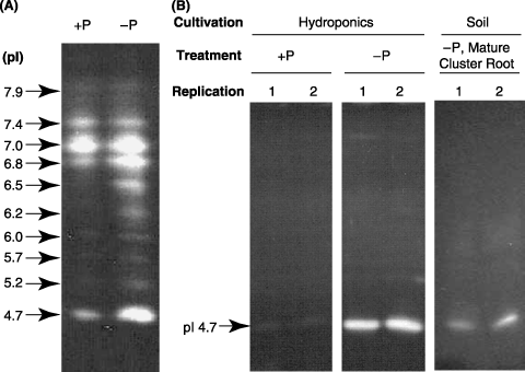
Figure 1  Activity staining of APase separated by isoelectric focusing. (A) Profile of APase isoforms in crude protein extracted from roots of hydroponically cultured white lupin. (B) Profile of secreted crude protein from roots of hydroponically cultured white lupin and extracted protein of rhizosphere cluster roots cultured in soil. pI, xxxx.

Figure 2  Visualization of the histochemical activity of APase for normal roots using ELF97 phosphate as a substrate. Hydroponically cultured white lupin roots were used for this experiment. Fluorescent precipitates of ELF97 (green color), the product of phosphatase activities, were observed using a fluorescence microscope. The results of the activity staining and autofluorescence are shown in the left and right panels, respectively. Upper and lower panels indicate the transverse sections of normal roots under +P and –P conditions, respectively.
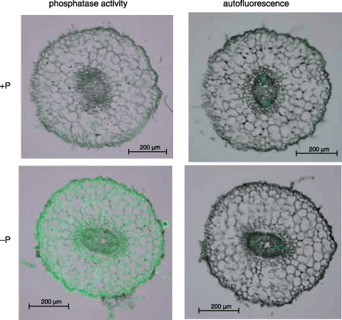
Figure 2  Visualization of the histochemical activity of APase for normal roots using ELF97 phosphate as a substrate. Hydroponically cultured white lupin roots were used for this experiment. Fluorescent precipitates of ELF97 (green color), the product of phosphatase activities, were observed using a fluorescence microscope. The results of the activity staining and autofluorescence are shown in the left and right panels, respectively. Upper and lower panels indicate the transverse sections of normal roots under +P and –P conditions, respectively.

Figure 3  Visualization of the histochemical activity of APase for cluster rootlets using ELF97 phosphate as a substrate. Hydroponically cultured white lupin roots were used for this experiment. The results of the activity staining and autofluorescence are shown in the left and right panels, respectively. Upper and lower panels indicate the cluster rootlets formed under +P and –P conditions, respectively.
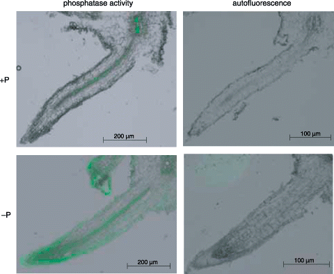
Figure 3  Visualization of the histochemical activity of APase for cluster rootlets using ELF97 phosphate as a substrate. Hydroponically cultured white lupin roots were used for this experiment. The results of the activity staining and autofluorescence are shown in the left and right panels, respectively. Upper and lower panels indicate the cluster rootlets formed under +P and –P conditions, respectively.

Figure 4  Visualization of the histochemical activity of APase for cluster rootlets formed under –P conditions using ELF97 phosphate as a substrate. Hydroponically and soil-cultured white lupin roots were used for this experiment. The results of the activity staining and autofluorescence are shown in the left (A,C,E) and right (B,D,F) panels, respectively. (A,B) Vertical sections of a cluster rootlet in a hydroponic plant; (C,D) transverse sections of a cluster rootlet in a hydroponic plant; (E,F) transverse sections of a cluster rootlet in a soil culture plant. Triangles indicate typical root hairs.
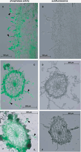
Figure 4  Visualization of the histochemical activity of APase for cluster rootlets formed under –P conditions using ELF97 phosphate as a substrate. Hydroponically and soil-cultured white lupin roots were used for this experiment. The results of the activity staining and autofluorescence are shown in the left (A,C,E) and right (B,D,F) panels, respectively. (A,B) Vertical sections of a cluster rootlet in a hydroponic plant; (C,D) transverse sections of a cluster rootlet in a hydroponic plant; (E,F) transverse sections of a cluster rootlet in a soil culture plant. Triangles indicate typical root hairs.

Reprints and Corporate Permissions

Please note: Selecting permissions does not provide access to the full text of the article, please see our help page How do I view content?

To request a reprint or corporate permissions for this article, please click on the relevant link below:

Academic Permissions

Please note: Selecting permissions does not provide access to the full text of the article, please see our help page How do I view content?

Obtain permissions instantly via Rightslink by clicking on the button below:

If you are unable to obtain permissions via Rightslink, please complete and submit this Permissions form. For more information, please visit our Permissions help page.