Figures & data
Table 1. Clinical features in Waldenström macroglobulinemia
Table 2. Cytogenetic and fluorescence in situ hybridization of del(20q) in Waldenström macroglobulinemia
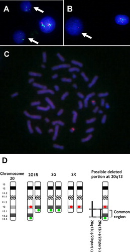
Figure 1. (A) An interphase cell with two green signals (pattern B: arrows) obtained from UPN 9. (B) An interphase cell with one red signals (pattern C: arrow) obtained from UPN 4. (C) A metaphase cell with two red signals on double del(20q) chromosomes without the presence of green signal obtained from UPN 4. (D) Schematic interpretation by FISH study on chromosome 20. The far-right portion indicates chromosome 20 with possible common deleted region in Waldenström macroglobulinemia. Solid vertical lines show possible deleted chromosome regions. 20q12(−)/20qter(+) indicates interstitial deletion of 20q12 but retention of 20qter: 20q12(+)/20qter(−) indicates deletion of 20qter but retention of the 20q12 locus. The region indicates the constant deleted region from distal to the 20q12 locus to proximal to the 20qter locus in four patients (UPNs 1, 4, 8, and 9).