Figures & data
Figure 1. A panel of photomicrographs depicting various non-Hodgkin lymphomas according to the Working Formulation: (a) malignant lymphoma (ML) small lymphocytic (H&E, ×100); (b) ML follicular predominantly small cleaved cells (H&E, ×100); (c) ML follicular predominantly large cells (H&E, ×100); (d) ML diffuse large cell (H&E, ×100); (e) ML lymphoblastic (H&E, ×100); and (f) ML diffuse mixed small and large cells (H&E, ×100).
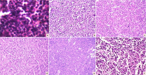
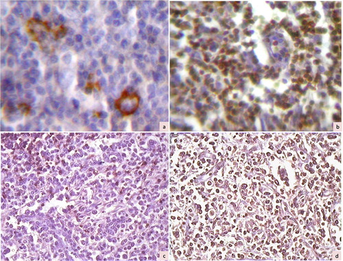
Figure 2. Immunohistochemical photomicrographs showing a case of T cell-rich B-cell lymphoma with large cells positive for CD20 (a, ×200) and small lymphocytes in the background staining for CD45RO (b, ×200). Another case of lymphoma (diffuse mixed small and large by Working Formulation) showing negative staining for CD20 (c, ×100) and diffuse positive staining for CD45RO (d, ×100), suggestive of T-cell lymphoma.