Figures & data
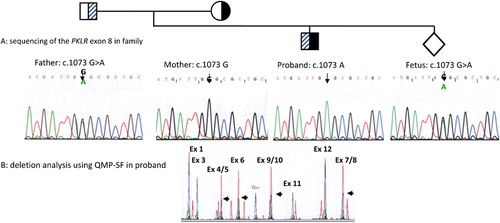
Figure 1. Genetic analysis of proband, parents and prenatal sample. (A) PCR-sequencing of exon 8 shows that the father is heterozygous for the PKLR: c.1073G>A mutation. Mother is hemizygous for normal exon 8 sequence, while the proband is hemizygous for the PKLR: c.1073G>A mutation. The fetus is heterozygous for the PKLR: c.1073G>A mutation. (B) Detection of the c.283+1914_c.1434del5006 deletion. A semi-quantitative PCR is performed with eight sets of primers which cover the whole PKLR gene. A fragment located outside of chromosome 1 is used as internal control for normalization. Chromatogram of a normal sample (red) is superimposed on that of the test sample (blue). Comparison of peak heights between test and normal demonstrates a deletion removing exon 4 to exon 10 in the proband (black arrows).