Figures & data
Figure 1. (A) Jurkat cells and (B) K562.
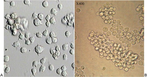
Figure 2. Morpholino delivery by Endo-Porter system. The fluorescence intensity and scattered pattern into cytosol reflecting suitable delivery can be seen.
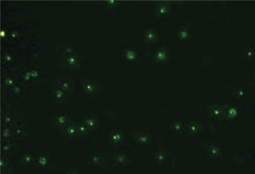
Figure 3. Flow cytometry analysis of K562 determines cell apoptosis exposed to Morpholino Oligo Antisense after (A) 24 hours, (B) 48 hours and (C) flow cytometry analysis of Jurkat cells: apoptosis rate decreased with increasing Morpholino concentration.
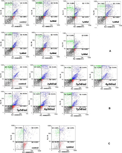
Figure 4. The relationship between different concentrations of Morpholino and cell apoptosis: all concentrations of Morpholino resulted in increasing cell apoptosis in comparison with negative group.
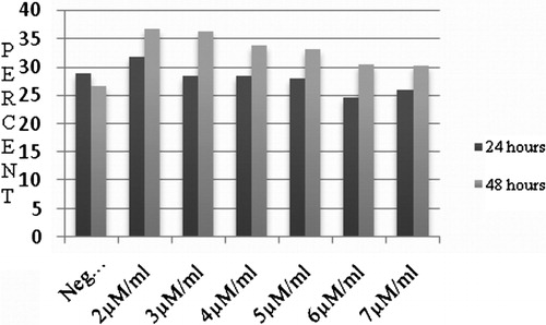
Figure 5. Effect of 2 μM/ml Morpholino on the cell viability in Ph(+) K562: the rate of cell viability significantly decreased in comparison with non-treated K562 cells (P⩽0·01).
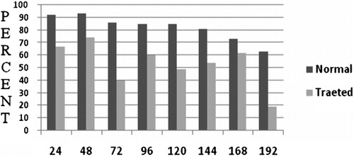
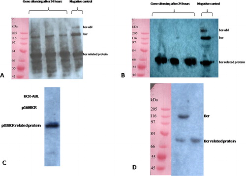
Figure 6. (A) Western blotting 24 hours following 2 μM/ml Morpholino Oligo Antisens treatment showing that p210bcr-abl suppressed and the level of p160bcr became weaker, (B) by 48 hours, bond of p210bcr-abl and p160bcr was not observed. The expression of p83bcr-related protein was considered as internal control group. (C) In the case of K562 treated with 5 μM/ml Morpholino, no p210bcr-abl and p160bcr band was detected after 24 hours. (D) Jurkat, the control group included cells treated with 2 μM/ml Morpholino.