Figures & data
Figure 1. Fibrin network prepared by adding thrombin to platelet rich plasma. Thick, white arrow = major, thick fibers; Thin, white arrow = minor, thin fibers. Scale = 1 μm. Insert: platelet with thin, white arrow indicating open canalicular membrane pore; thick white arrow = major fibers leaving platelet. Scale = 1 μm.
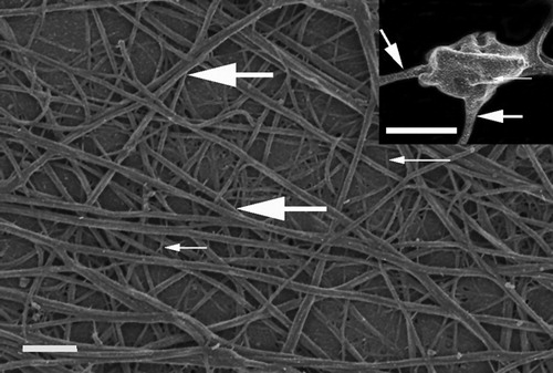
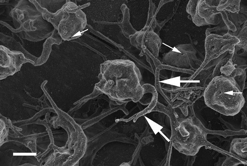
Figure 2. Platelet rich plasma from a citrated whole blood sample recalcified with addition of 0·2M CaCl2. Thick, white arrows = fibrin fibers associated with platelets; thin, white arrows = open canalicular membrane pores. Scale = 1 μm.
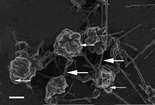
Figure 3. Platelet rich plasma from citrated whole blood sample, without recalcifying or addition of thrombin. Thick, white arrows = fibrin fibers associated with platelets; thin, white arrows = open canalicular membrane pores. Scale = 1 μm.