Figures & data
Table 1. Oligonucleotide sequences of shRNAs
Figure 1. Results of 1% agarose gel electrophoresis. (A) A single electrophoretic band smaller than 100 bp by 1% agarose gel electrophoresis indicated the success of Oligo single strand annealing; (B) shRNA expression vectors targeting BAALC could be digested by BamH I and Pst I.
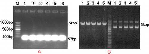
Figure 2. Effects of BAALC shRNA on mRNA and protein expression. (A) Results of 1% agarose gel electrophoresis showed that BAALC mRNA expression was decreased in cells transfected with pGPU6-BAALC-shRNA1-4. However, no change was observed in cells transfected with pGPU6-shRNA-NC. Lane 1: control; Lane 2: GAPDH-shRNA; Lane 3: GAPHD-shRNA-NC; BAALC: Lane 3: BAALC-shRNA-NC; Lanes 4–7: BAALC-shRNA-1-4. (B) Results of western blot showed that BAALC protein levels were markedly inhibited in pGPU6-BAALC-shRNA1-4 transfected leukaemia cells, in contrast to the negative control plasmid-transfected cells. Lane 1: BAALC-shRNA-NC, Lanes 2–5: BAALC-shRNA-1-4.
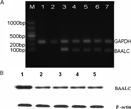
Figure 3. Mean ODs were measured by CCK-8 assay, proliferation of KG1a-BAALC shRNA cells was significantly suppressed at each different time point (48, 72 and 96 hours), compared with the control groups during the time course.
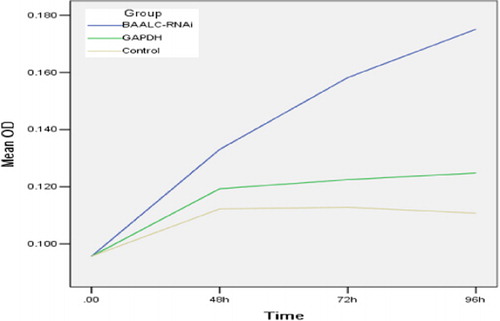
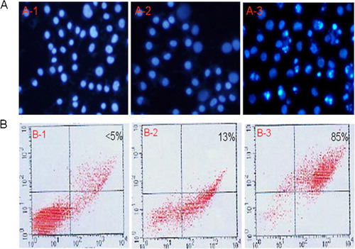
Figure 4. Effects of BAALC shRNA on apoptosis in KG1a cells. (A) KG1a cells were stained with DAPI and observed under fluorescence microscopy (punctuated, granular, and brighter nuclei). (B) Results of Annexin V-FITC (measured by FACS).