Figures & data
Table 1. Diazinon toxicity impacts on body and organ weight
Table 2. Effects of diazinon on testicle antioxidants and pro-oxidant (lipid peroxidation) level
Table 3. The influence of diazinon on serum testosterone synthesis and lactate dehydrogenase activity
Figure 1. Photomicrographs of testis sections of DZN-treated animals (1 week), (A) normal seminiferous tubules (L indicates lumen) in untreated animals, (B) shrinkage (*) of seminiferous tubules in 10 mg/kg b.w. DZN-treated group, (C) halo appearance or vacuoles (arrows) in the seminiferous tubules of 15 mg/kg b.w. DZN-treated group, and (D) more vacuoles (arrows) in the seminiferous tubules of 30 mg/kg b.w. DZN-treated group. Vacuoles were also observed in 2 weeks of DZN exposure – 10, 15, and 30 mg/kg groups. Slides were stained with haematoxylin-eosin dye. Scale, 200 µm.
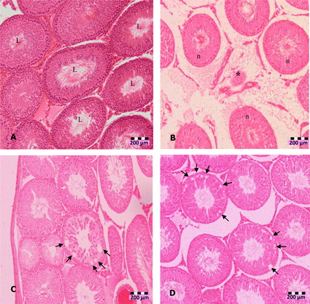
Figure 2. Photomicrographs of testis sections of DZN-treated animals, (A) in 2 weeks of DZN exposure (15 and 30 mg/kg b.w.), treated animals showed disorganization and disintegration of cell, which caused secondary spermatocyte (arrows) moved into lumen (100×), (B) shrinkage (*) of seminiferous tubules and loss of cells (lc), (C) more immature cells appeared in the lumen of 30 mg/kg b.w. DZN-treated animals (arrows), and (D) abnormal formation of spermatozoa in the lumen. Slides were stained with haematoxylin-eosin dye. Scale of (A) and (B) – 200 µm, (C) and (D) – 100 µm.
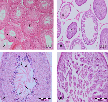
Table 4. Diazinon induced gonadotoxicity effects
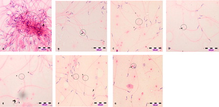
Figure 3. Photomicrographs show abnormal sperms, (A) clumping, (B) head deformity (arrow), (C) hookless (a) and coiled tail (b), (D) bent-tail (arrow, (E) broken-head (a) neck's defect (n), (F) broken head (a), broken tail (b) and bent tail (arrow), (G) double head (arrow). Slides were stained with haematoxylin-eosin dye. Scale, 100 µm.