Figures & data
Table 1 Experimental study protocol
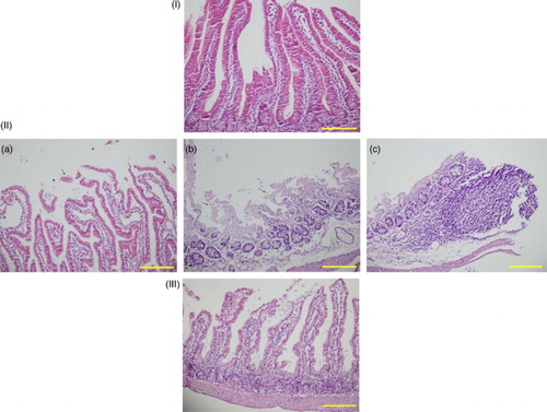
Figure 1 Sections of the small intestine were stained with hematoxylin–eosin, scale bar = 200 μm. (I) Normal histological appearance of the small intestine tissues in the control rats. (II) Histopathological effects of high dose methotrexate on the small intestine structure. (a) Degeneration of surface and crypt epithelium. (b) Degeneration of villus structure. (c) Inflammatory cell infiltration. (III) Section indicating that lycopene treatment in the Mtx-L group is providing significant histological improvement.