Figures & data
Table 1. Effect catecholamines on genes and cytokines-related HSCs’ mobilization
Figure 1. Epinephrine regulation of CXCR4 mRNA synthesis; HSCs were cultured for 5 hours with 10 µM epinephrine. CXCR4 mRNA levels were measured by quantitative PCR. The results reflect the mean of at least three experiments; the error bars reflect SD of the mean (**P < 0.01).
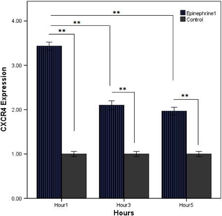
Figure 2. Effect of G-CSF and epinephrine on CXCR4 expression level; CXCR4 mRNA was measured by quantitative PCR. (A) Cord blood HSCs were cultured for 1–5 hours in the presence of G-CSF (100 ng/ml) plus epinephrine (10 µM). (B) Comparing effect of alone epinephrine (10 µM) with epinephrine (10 µM) plus G-CSF (100 ng/ml) on CXCR4 expression of HSCs. The results reflect the means of triplicate determinations; the error bars represent SD of the mean. *P < 0.05, **P < 0.01.
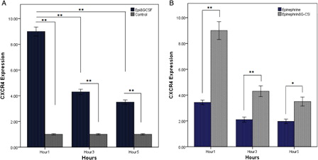
Figure 3. The direct role of beta-adrenergic receptors on expression of CXCR4. (A) Quantitative RT-PCR analysis of CXCR4 mRNA in HSCs for 5 hours of incubation with G-CSF (100 ng/ml) and epinephrine (10 µM) plus propranolol (1 µM). (B) Cord blood HSCs were incubated with isoproterenol (10 µM) for 5 hours. The results are expressed as mean (±SD) of triplicate cultures. (C) Levels of CXCR4 mRNA were measured by quantitative PCR. G-CSF (100 ng/mL) plus isoproterenol (10 µM) increases the expression of CXCR4 in CD34+ HSCs. (D) Studying the effect of alone isoproterenol (10 µM) with isoproterenol (10 µM) plus G-CSF (100 ng/ml) on CXCR4 expression of HSCs. The results reflect the mean fold (±SD) mRNA change relative to cells cultured as a control. *P < 0.05, **P < 0.01.
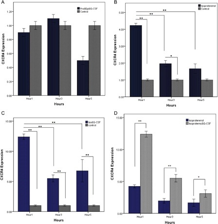
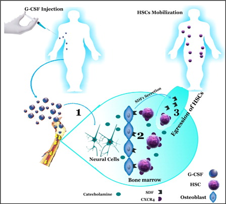
Figure 4. Mobilization of hematopoietic stem cell induced by G-CSF and catecholamines (1) G-CSF-induced up-regulation of catecholamines from the sympathetic neuron. (2) Catecholamines effect on CXCR4 and SDF-1 expression. (3) Expression of SDF-1 is reduced in BM environment and is secreted into circulation, and CXCR4 expression in hematopoietic is increased inducing egression of HSC from BM. SDF-1, stromal-derived factor-1; CXCR4, CXC motif, receptor 4; HSC, hematopoietic stem cell; G-CSF, granulocyte colony-stimulating factor.