Figures & data
Table 1. Demographic and hematological profile of patients with prominent splenomegaly without lymphadenopathy
Figure 1. The categories of mature lymphoid neoplasms, their frequencies and the diagnostic method utilized for their immunophenotyping in patients with prominent splenomegaly without lymphadenopathy.
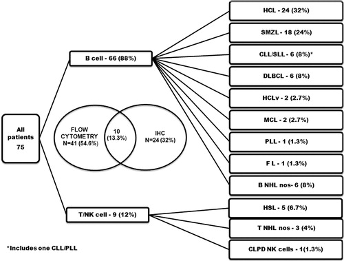
Figure 2. The morphological features of lymphoid cells in peripheral blood and/ or bone marrow aspirate (100x; May Grunwald Giemsa stain) (A) Hairy cells of HCL. (B) SMZL showing monocytoid lymphocytes. Short polar villi seen in one of them (C) Villous lymphocyte of SMZL (D) CLL showing small lymphocytes along with occasional prolymphocytes (E) B-cell PLL showing predominantly prolymphocytes (F) MCL showing lymphoid cells with irregular cleaved nucleus (G) Intravascular large B cell lymphoma showing blastoid appearing lymphoid cells (H) γδ HSTL showing medium to large sized lymphoid cells (I) CLPD NK cells showing large granular lymphocytes.
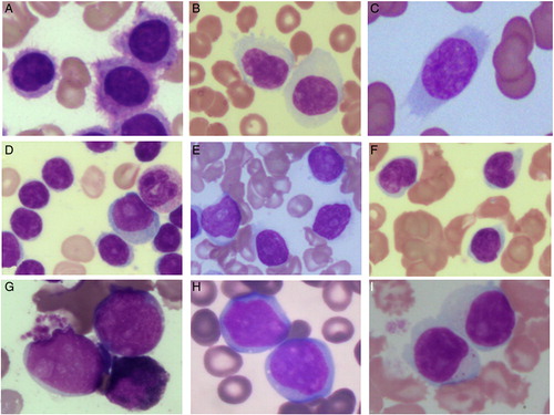
Figure 3. The morphology and salient immunohistochemistry findings of mature lymphoid neoplasms in bone marrow trephine biopsy. HCL showing typical ‘fried egg’ appearance (A) [Haematoxylin and eosin (H&E), 40x] and cytoplasmic positivity for CD72 (DBA.44) (B) [IHC, 40x]. MCL showing diffuse infiltration of bone marrow (C) [H&E, 40x] with nuclear positivity for cyclin D1 (D) [IHC, 40x]. SMZL showing nodular lymphoid infiltrate (E) [H&E, 10x] with intrasinusoidal and interstitial infiltrate highlighted by CD20 (F) [IHC, 20x]. DLBCL showing diffuse infiltration by large atypical lymphoid cells (G) [H&E, 40x] which are positive for CD20 (H) [IHC, 40x]. Intravascular large B cell lymphoma showing scattered atypical large cells (I) (H&E, 40x), the intrasinusoidal nature of which is highlighted by CD20 (J) [IHC, 40x]. Diffuse infiltrate of atypical large lymphoid cells with prominent eosinophilic nucleolus (K) [H&E, 40x], which are CD20+ CD5+ CD10− (not shown) and Cyclin D1− (L) [IHC, 40x] raising a differential diagnosis of cyclin D1 negative blastoid MCL and CD5+ DLBCL.
![Figure 3. The morphology and salient immunohistochemistry findings of mature lymphoid neoplasms in bone marrow trephine biopsy. HCL showing typical ‘fried egg’ appearance (A) [Haematoxylin and eosin (H&E), 40x] and cytoplasmic positivity for CD72 (DBA.44) (B) [IHC, 40x]. MCL showing diffuse infiltration of bone marrow (C) [H&E, 40x] with nuclear positivity for cyclin D1 (D) [IHC, 40x]. SMZL showing nodular lymphoid infiltrate (E) [H&E, 10x] with intrasinusoidal and interstitial infiltrate highlighted by CD20 (F) [IHC, 20x]. DLBCL showing diffuse infiltration by large atypical lymphoid cells (G) [H&E, 40x] which are positive for CD20 (H) [IHC, 40x]. Intravascular large B cell lymphoma showing scattered atypical large cells (I) (H&E, 40x), the intrasinusoidal nature of which is highlighted by CD20 (J) [IHC, 40x]. Diffuse infiltrate of atypical large lymphoid cells with prominent eosinophilic nucleolus (K) [H&E, 40x], which are CD20+ CD5+ CD10− (not shown) and Cyclin D1− (L) [IHC, 40x] raising a differential diagnosis of cyclin D1 negative blastoid MCL and CD5+ DLBCL.](/cms/asset/3f9eb6f6-fc31-40e4-a544-6bedec127743/yhem_a_11659347_f0003_b.jpg)
Table 2. Summary of clinical and hematological findings of mature lymphoid neoplasms diagnosed in patients with prominent splenomegaly without lymphadenopathy
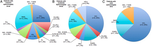
Figure 4. The distribution of mature lymphoid neoplasms in patients presenting with massive (A), moderate (B), and mild (C) splenomegaly.