Figures & data
Figure 1. In vivo human RPE and choroidal GAPDH and nitrotyrosine. Ischemic choroid and RPE (A) demonstrate GAPDH nuclear translocation in RPE and vascular endothelial cells (C) as well as considerable immunoreactive nitrotyrosine (E). RPE and choroid of control eye show only cytoplasmic GAPDH and minimal nitrotyrosine (B, D, F). Bar = 25 µm.
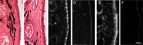
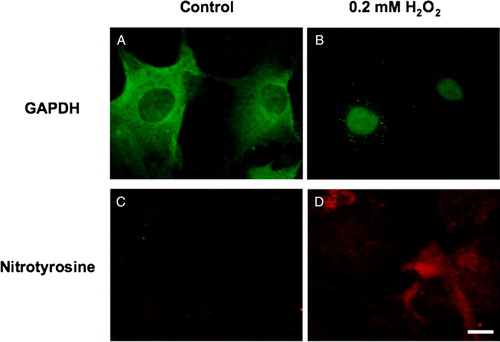
Figure 2. In vitro human RPE GAPDH and nitrotyrosine. Cultured human RPE cells (second passage) were exposed to control medium (A, C) or medium containing 200 µM hydrogen peroxide (B, D) for 24 hours. Immunocytochemistry for GAPDH (A, B) and nitrotyrosine (C, D) was performed. Bar = 10 µm.