Figures & data
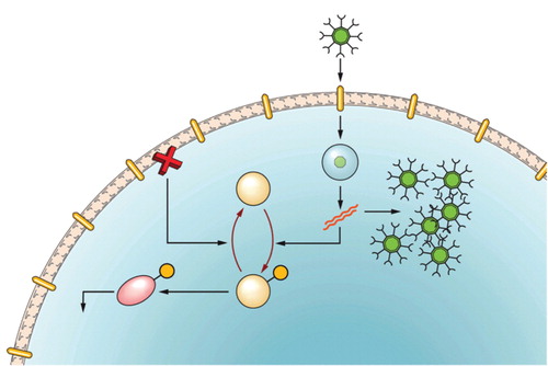
Figure 1. A brief overview of the infective cycle of reovirus. 1. Reovirus binds JAM-1 and sialic acid receptors ubiquitously found on the cell surface. 2. Reovirus enters the cell by endocytosis with subsequent cleavage of sigma and mu proteins from the outer shell of the virus to form an intermediate sub viral particle (ISVP). This is released into the cytoplasm. 3. Viral dsRNA is copied within the viral core and released. 4. In normal cells expressing wild-type Ras, dsRNA activates PKR by phosphorylation. 5. Phosphorylated PKR subsequently activates eIF2α, which halts viral protein synthesis, resulting in failed infection. 6. In contrast, in cells with an activated/mutated Ras pathway, PKR is held in an inactive, hypophosphorylated state. 7. In these cells viral infection ensues. Viral proteins are produced using the cells machinery, viral factories form in the cytoplasm and reoviral particles assemble.