2,025
Views
44
CrossRef citations to date
0
Altmetric
Reviews

The use of the NIS reporter gene for optimizing oncolytic virotherapy

&

Figures & data

Figure 1. Representative whole body nuclear imaging of mice infected with oncolytic virus expressing NIS following NIS-mediated uptake of radiotracer. The thyroid (T), stomach (S) and bladder (B) can be detected due to endogenous NIS expression and radiotracer excretion. Low-resolution SPECT/CT imaging clearly identifies intratumoral radiotracer uptake. High-resolution SPECT/CT imaging allows for spatial resolution of intratumoral infected centers.

Figure 1. Representative whole body nuclear imaging of mice infected with oncolytic virus expressing NIS following NIS-mediated uptake of radiotracer. The thyroid (T), stomach (S) and bladder (B) can be detected due to endogenous NIS expression and radiotracer excretion. Low-resolution SPECT/CT imaging clearly identifies intratumoral radiotracer uptake. High-resolution SPECT/CT imaging allows for spatial resolution of intratumoral infected centers.

Table 1. Available radiotracers and corresponding half-lives, decay mode and major γ emission energy for NIS-mediated imaging with SPECT of PET instrumentation.

Figure 2. Schematic of NIS expression at the cellular level and corresponding NIS-mediated radiotracer uptake detected and quantified with nuclear imaging techniques. A. Tumor cell does not naturally express NIS and therefore does not concentrate radiotracers. B. Upon viral infection, the viral genome (green) will be replicated and viral genes, along with transgenes (blue), will be transcribed and translated into functional proteins. Expression of the NIS transgene (blue) leads to production of NIS protein that allows radiotracer uptake, corresponding with nuclear imaging detection. C. As viral replication continues, there is increased expression of viral genes and transgenes, leading to increased NIS protein for increased radiotracer uptake detected by nuclear imaging. D. As viral proteins continue to be produced, viral particles are assembled and exit the cell through budding or lysis. Cytopathic effects on cell cause loss of radiotracer uptake capabilities, corresponding to decrease in nuclear imaging detection.

Figure 2. Schematic of NIS expression at the cellular level and corresponding NIS-mediated radiotracer uptake detected and quantified with nuclear imaging techniques. A. Tumor cell does not naturally express NIS and therefore does not concentrate radiotracers. B. Upon viral infection, the viral genome (green) will be replicated and viral genes, along with transgenes (blue), will be transcribed and translated into functional proteins. Expression of the NIS transgene (blue) leads to production of NIS protein that allows radiotracer uptake, corresponding with nuclear imaging detection. C. As viral replication continues, there is increased expression of viral genes and transgenes, leading to increased NIS protein for increased radiotracer uptake detected by nuclear imaging. D. As viral proteins continue to be produced, viral particles are assembled and exit the cell through budding or lysis. Cytopathic effects on cell cause loss of radiotracer uptake capabilities, corresponding to decrease in nuclear imaging detection.

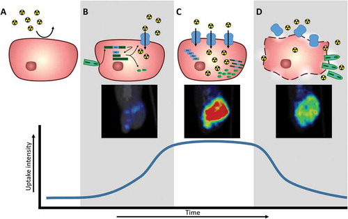
Table 2. Table of oncolytic viruses expressing the NIS transgene to mediate radiotracer uptake for in vivo nuclear imaging monitoring of infection, separated by oncolytic virus and tumor type.**

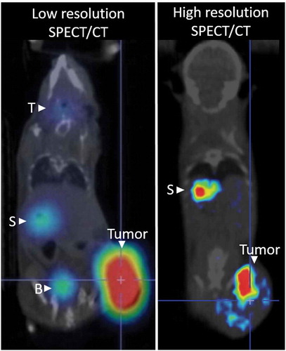
Table 3. Resolution and sensitivity of imaging instruments used to detect oncolytic virus replication via NIS mediated radiotracer uptake.