1,248
Views
20
CrossRef citations to date
0
Altmetric
Reviews

Tissue slide-based microRNA characterization of tumors: how detailed could diagnosis become for cancer medicine?

Figures & data

Table 1. Variations and evolution of tissue slide-based assays for in situ detection of miRNAs.

Table 2. Potential clinical applications of miRNA-based in situ hybridization assays.

Figure 1. Automated platform for high-content marker analysis of tumor tissues. (A) Key components of in situ miRNA detection assay. (B) Purposes of key components and the steps they take part in, from tissue digestion to covalently linked deposition of fluorescent labels. (C) Virtual staining rendition of a 9plex assay that co-detects three RNA markers (two miRNAs + control rRNA) and six protein markers (three cell type-specific + three functional/prognostic markers); black dots indicate nuclei revealed by counterstaining with DAPI. Top left, representative miR-21 stain (green) on an ERPRHER2 breast cancer case in the first round of HRP-mediated deposition of reactive substrate conjugated with a fluorochrome. Sequential rounds of HRP-mediated deposition of substrates conjugated with spectrally unique fluorochromes are carried out to detect probes against other RNA markers with appropriate anti-hapten primary and secondary antibody combinations or protein markers with appropriate primary and secondary antibody combinations. Each new staining round requires a peroxide block step to inactive HRP from the preceding round.

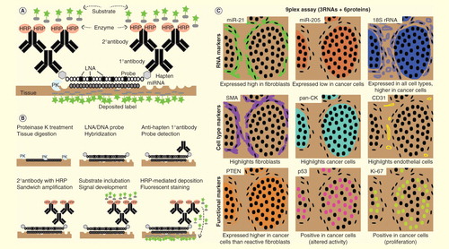
Figure 1. Automated platform for high-content marker analysis of tumor tissues. (A) Key components of in situ miRNA detection assay. (B) Purposes of key components and the steps they take part in, from tissue digestion to covalently linked deposition of fluorescent labels. (C) Virtual staining rendition of a 9plex assay that co-detects three RNA markers (two miRNAs + control rRNA) and six protein markers (three cell type-specific + three functional/prognostic markers); black dots indicate nuclei revealed by counterstaining with DAPI. Top left, representative miR-21 stain (green) on an ER−PR−HER2− breast cancer case in the first round of HRP-mediated deposition of reactive substrate conjugated with a fluorochrome. Sequential rounds of HRP-mediated deposition of substrates conjugated with spectrally unique fluorochromes are carried out to detect probes against other RNA markers with appropriate anti-hapten primary and secondary antibody combinations or protein markers with appropriate primary and secondary antibody combinations. Each new staining round requires a peroxide block step to inactive HRP from the preceding round.