Figures & data
Figure 1. Structure of an antibody and its associated Fc-mediated effector functions that can be activated by the Fc region. The Fab region is responsible for specific antigen binding whereas the Fc region binds to FcγRs in immune cells, resulting in phagocytosis, cytokine release, ADCC and CDC.

Figure 2. Different FcγRs present in myeloid cells. Unlike FcγRII, FcγRI and FcγRIII require the common gamma chain for ITAM-mediated signaling. FcγRI binds strongly to IgG while FcγRII and FcγRIII bind strongly to immune complexes but not monomeric IgG. FcγRIIB is an inhibitory receptor bearing ITIM that counteracts ITAM-mediated signaling. FcRn can recycle IgG as it binds strongly to IgG at lower pH, resulting in increased half-life of antibodies in blood.
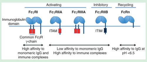
Figure 3. The different FcγRs involved in virus neutralization at various antibody concentrations. At high antibody concentrations, immune aggregates formed can coligate the lowly expressed inhibitory FcγRIIB to inhibit FcγR-mediated phagocytosis. Small sized immune complexes, in contrast, utilize the activating FcγRs signaling for phagocytosis.
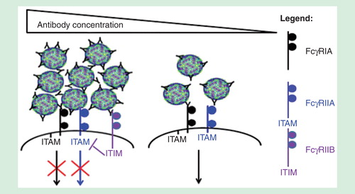
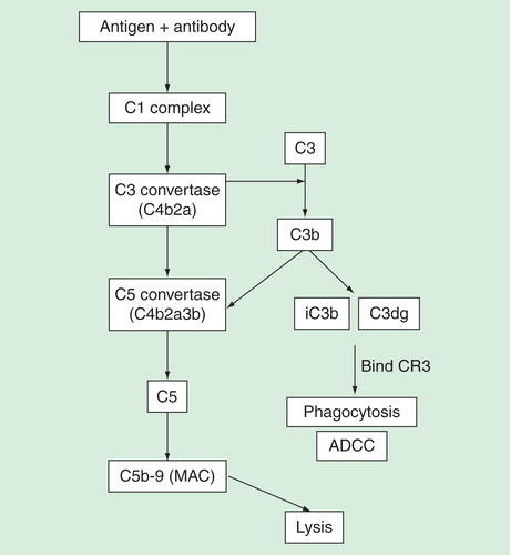
Figure 4. Immune complexes can trigger the complement pathway to result in either eventual lysis or phagocytosis of target antigen. C1q binding to immune complexes results in formation of C3 and C5 convertases that cleave C3 and C5, respectively. Cleavage of C3 results in fragments iC3b and C3dg that binds to CR3 present in leukocytes or NK-cells, resulting in phagocytosis and ADCC. C5 cleavage, on the other hand, results in formation of MAC that forms membrane pores and eventual lysis of target. Phagocytosis, ADCC and lysis assays can be used to assess involvement of complements.