
Figures & data
Figure 1. The RNS® neurostimulator and NeuroPace® leads.
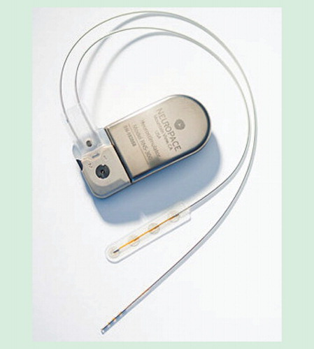
Figure 2. The RNS System. The implantable components of the RNS System include a neurostimulator and depth and cortical strip leads. The programmer is a laptop computer with proprietary software and custom telemetry components that the physician uses to communicate with the neurostimulator. The programmer can configure neurostimulator settings and retrieve data stored on the neurostimulator. The remote monitor is a home-use monitoring device used by the patient to retrieve data stored on the neurostimulator. The PDMS is a centralized database, which contains data uploaded from the programmer and remote monitor. Neurostimulator data and detection settings can also be transferred from PDMS to the programmer.
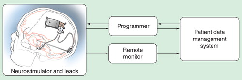
Figure 3. Example of a four-channel ECoG segment stored by the neurostimulator as displayed on the PDMS. In this example, the neurostimulator has been programmed to detect paroxysmal high-amplitude sharply contoured discharges on channel 3, which is typical of this patient’s interictal epileptiform activity. ‘B2’ denotes detection of epileptiform activity on the third channel. ‘Tr’ indicates delivery of responsive stimulation. Following the initial detection, the electrocoticogram is displayed in blue. When responsive stimulation is delivered, and immediately afterward, there is an artifact in the recording.
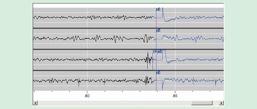
Figure 4. Skull x-rays of two patients implanted with the RNS® neurostimulator and NeuroPace® leads. The patient on the left is a female, with seizures arising bilaterally from the mesial temporal lobes. The x-ray shows NeuroPace depth leads implanted longitudinally in the left and right hippocampi. The Leads are connected to the RNS neurostimulator shown implanted in the parietal bone. The patient on the right is a male, with seizures arising from the left frontal lobe. The x-ray shows NeuroPace cortical strip leads implanted over the left frontal lateral cortex. The leads are connected to the RNS neurostimulator implanted in the parietal skull.
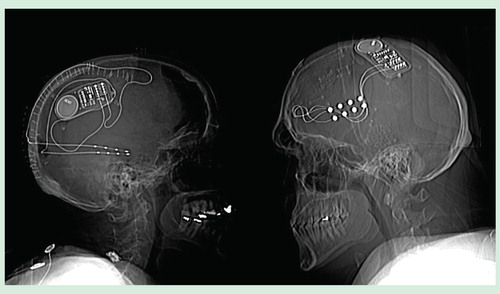
Figure 5. Responder rate and median percent reduction in seizure frequency during the open-label period in the pivotal study.
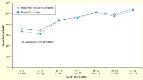
Table 1. Pivotal study – primary safety endpoint.