2,538
Views
1
CrossRef citations to date
0
Altmetric
Editorial

A simple guide to dermatoscopic diagnosis of melanocytic and nonmelanocytic skin lesions

Pages 333-339 | Published online: 10 Jan 2014

Figures & data

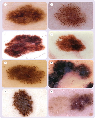
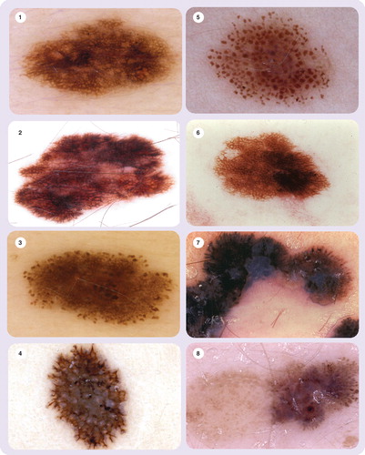
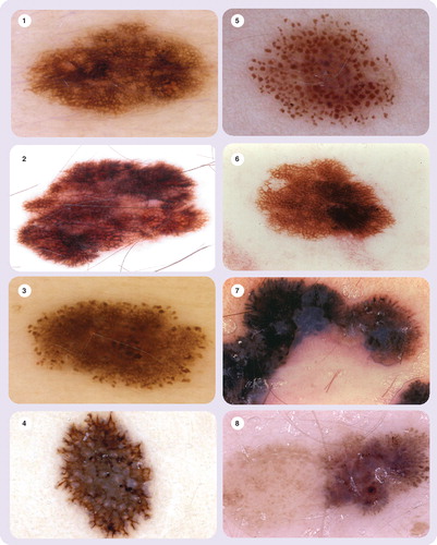
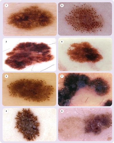
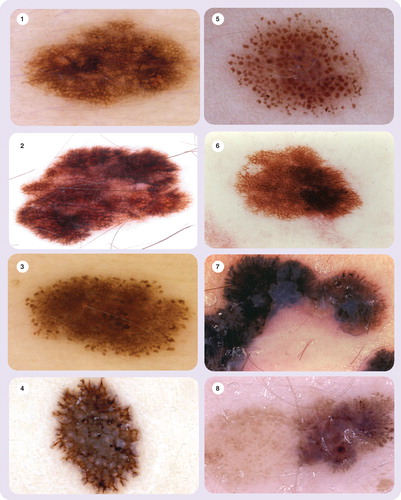
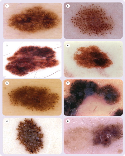
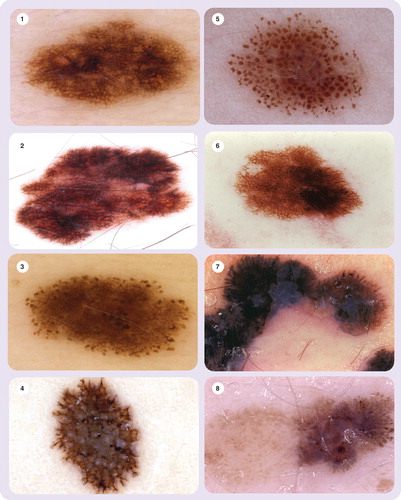
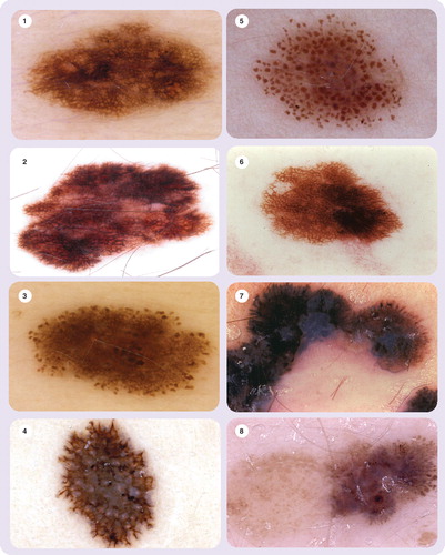
Figure 1. Pigment network
Figure 2. Pigment network
Figure 3. Aggregated globules and pigment network
Figure 4. Aggregated globules
Figure 5. Streaks (radial streaming)
Figure 6. Pseudopods and regression (white and blue areas)
Figure 7. Regular streaks
Figure 8. Streaks (pseudopods).
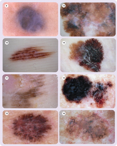
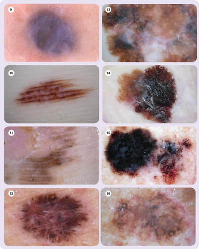
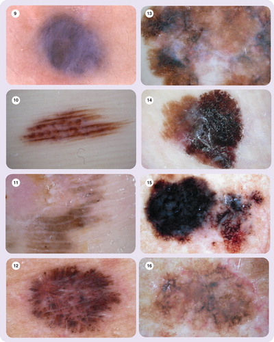
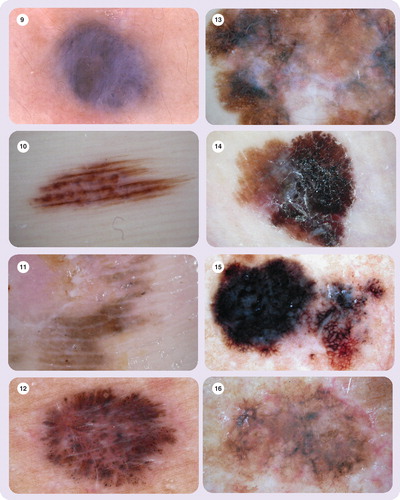
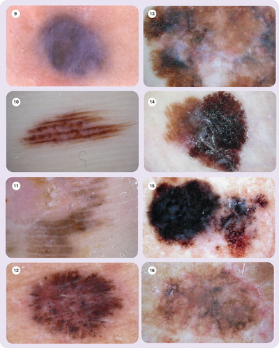
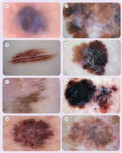
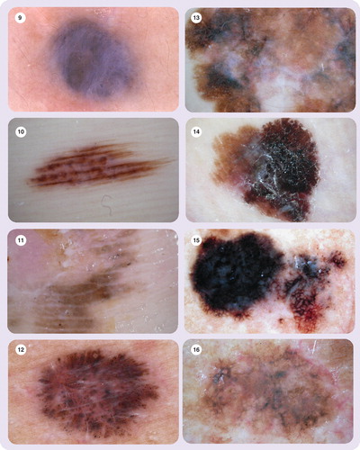
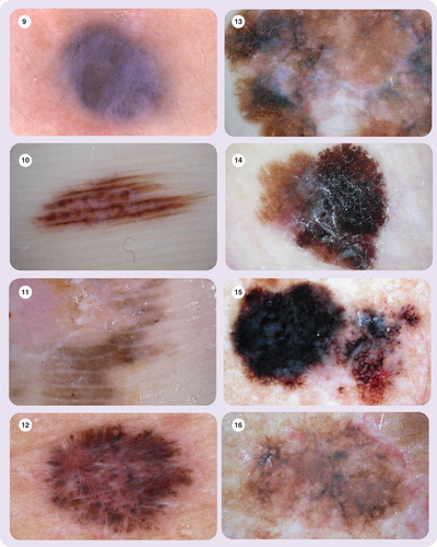
Figure 9. Homogeneous blue pigmentation
Figure 10. Parallel furrow pattern
Figure 11. Parallel ridge pattern
Figure 12. Dots
Figure 12. Dots
Figure 13. Regression structures (blue areas are melanosis, white areas are fibrosis)
Figure 14. Blue-whitish veil
Figure 15. Blotches, pigment network, streaks and regression
Figure 16. Hypopigmentation.
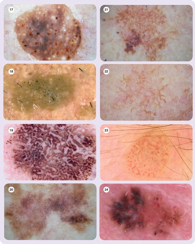
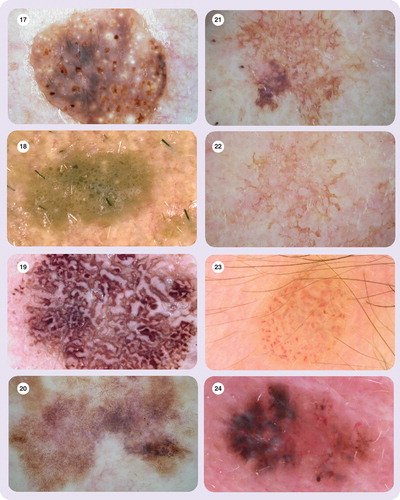
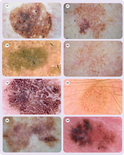
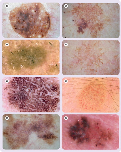
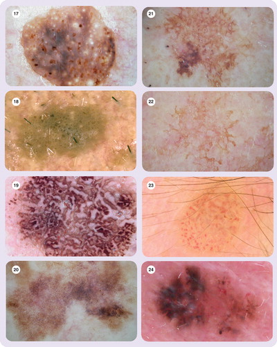
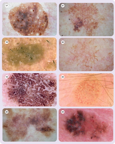
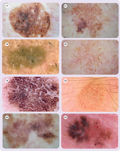
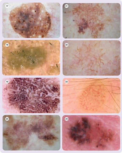
Figure 17. Multiple milia-like cysts
Figure 18. Comedo-like openings
Figure 19. Fissures/ridges with a brain-like appearance
Figure 20. Light brown fingerprint-like structures
Figure 21. Moth-eaten border
Figure 22. Jelly-like border
Figure 23. Vascular pattern with hairpin vessels
Figure 24. Leaf-like structures.
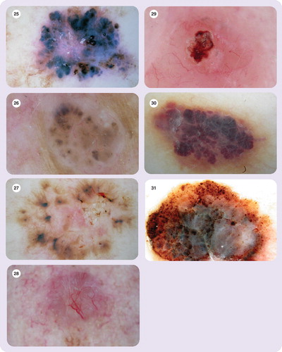
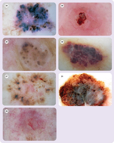
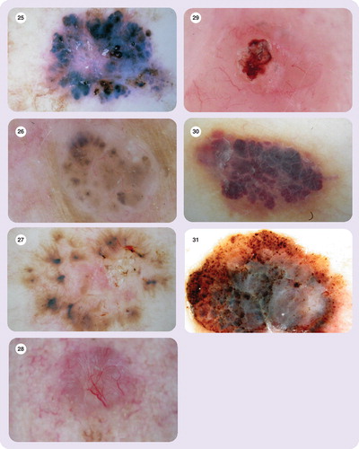
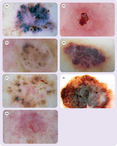
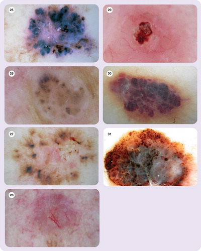
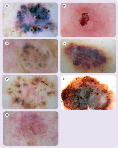
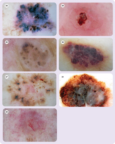
Figure 25. Large blue-gray ovoid nests
Figure 26. Multiple blue-gray globules
Figure 27. Spoke wheel areas
Figure 28. Vascular pattern with arborizing vessels
Figure 29. Ulceration
Figure 30. Lacunar pattern
Figure 31. Dots, globules, blue-whitish veil and streaks.

Reprints and Corporate Permissions

Please note: Selecting permissions does not provide access to the full text of the article, please see our help page How do I view content?

To request a reprint or corporate permissions for this article, please click on the relevant link below:

Academic Permissions

Please note: Selecting permissions does not provide access to the full text of the article, please see our help page How do I view content?

Obtain permissions instantly via Rightslink by clicking on the button below:

If you are unable to obtain permissions via Rightslink, please complete and submit this Permissions form. For more information, please visit our Permissions help page.