Figures & data
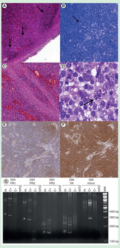
Figure 1. Piringer-Kuchinka lymphadenitis features. (A) Cortical follicles with zonal hyperplastic features and germinal centers showing ragged, ‘moth-eaten’ margins with tingible body macrophages, HE, 4×. (B) Clusters of epithelioid hystiocytes forming granulomatous aggregates, Giemsa, 4×. (C) Parasinusoidal monocytoid B-lymphocytes, characteristic hallmark of toxoplasma lymphadenitis, HE, 20×. (D) Parasitic cyst with multiple amastigotes, confirming the infectious etiology, HE, 100×. (E, F) Double positivity to CD20/CD5 markers in paracortical B-cells is an unusual finding in reactive condition, and may lead to a more careful investigation of the process, IHC, 4×. (G) Clonality assay: PCR amplification fragments indicative of patient-specific clonal IGH FR1, IGH FR2, IGH FR3 and IGK VK gene rearrangements which are indicated with an asterisk.