Figures & data
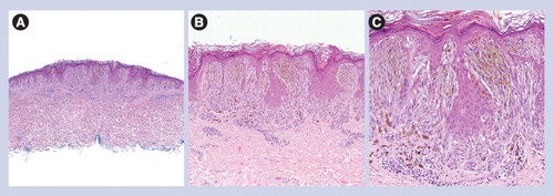
(A) Low power circumscription and symmetry with epidermal hyperplasia (40×). (B) Nests of epithelioid melanocytes are arranged vertically to the skin surface with ‘raining down’ appearance (100×). (C) Higher magnification shows epithelioid melanocytes, Kamino body and occasional pagetoid scattering (200×).
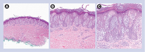
(A) Circumscribed and symmetric lesion (40×). (B) Mostly junctional nevus composed of heavily pigmented spindled melanocytes (100×). (C) Heavily pigmented and spindled melanocytes without cytologic atypia (200×).
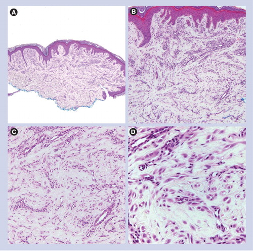
(A) Dermal-based lesion (40×). (B & C) Spindled and epithelioid melanocytes are arranged as fascicles or single units between sclerotic collagen bundles (100× and 200×, respectively). (D) Sclerotic collagen and epithelioid/spindled melanocytes (400×).
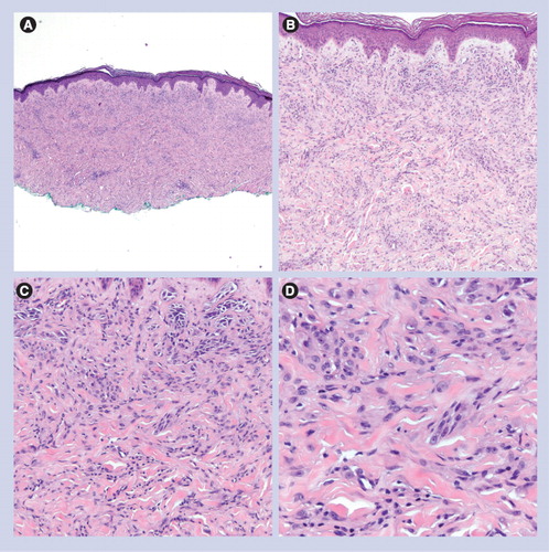
(A) Dermal-based lesion (40×). (B & C) Spindled and epithelioid melanocytes are set in a dense fibrous stroma, associated with prominent blood vessels (100× and 200×, respectively). (D) Epithelioid/spindled melanocytes and prominent vessels (400×).
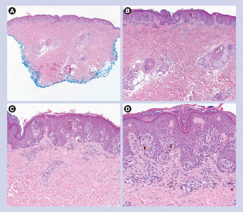
(A) Small and circumscribed lesion (20×). (B & C) Epidermal hyperplasia and bridging of junctional nests (40× and 100×, respectively). (D) Horizontal bridging of junctional nests with concentric fibroplasia and mild dermal inflammation. The melanocytes are epithelioid. Kamino bodies are present (200×).
(A) Dermal-based lesion with sheet-like growth pattern and incomplete maturation (40×). (B & C) Nests do not become smaller in deeper dermis (100× and 200×, respectively). (D) Melanocytes are epithelioid with nuclear pleomorphism (400×).
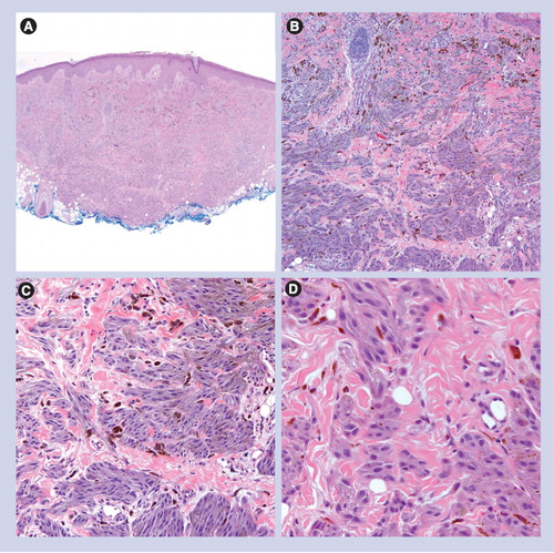
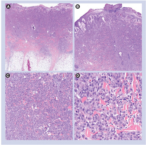
(A) Expansile dermal tumor with surface ulceration (20×). (B & C) Sheet-like growth pattern (40× and 100×, respectively). (D) Epithelioid melanocytes with nuclear pleomorphism, prominent nucleoli and mitoses (400×).