Figures & data
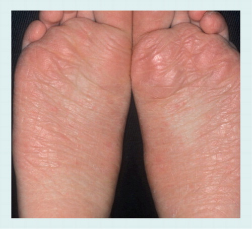
Mild erythema and scaling.
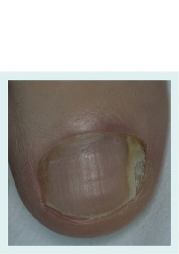
Differential diagnosis with distal subungual onychomycosis requires mycology.
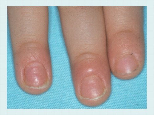
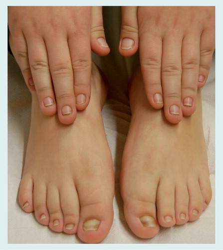
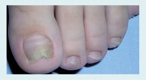
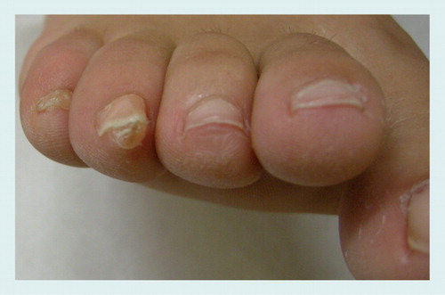
Nail lesions in a 7-year-old boy. All nails show mild thickening with distal onycholysis and subungual hyperkeratosis.
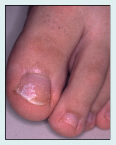
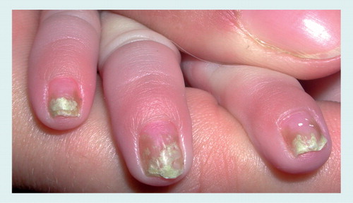
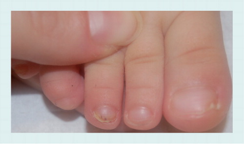
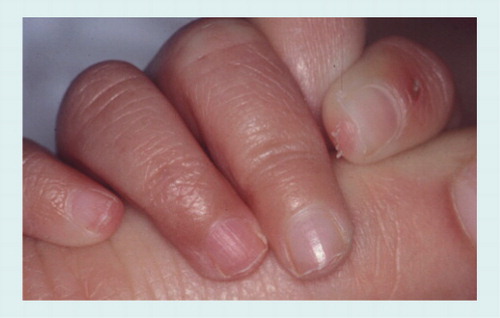
Diffuse crumbling of the nail plate.
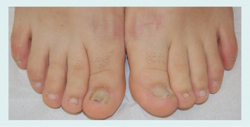
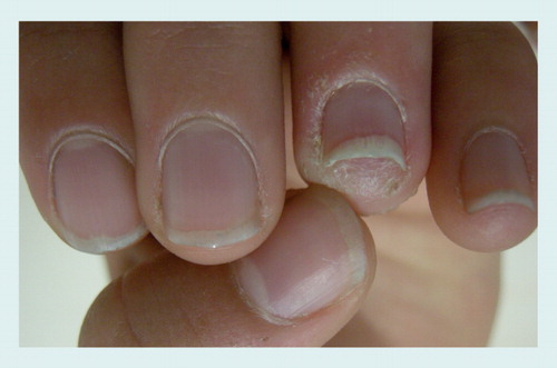
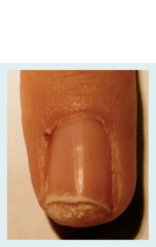
Onycholysis with erythematous border and splinter hemorrhages.
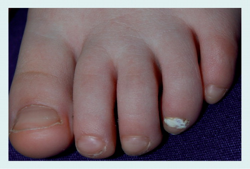
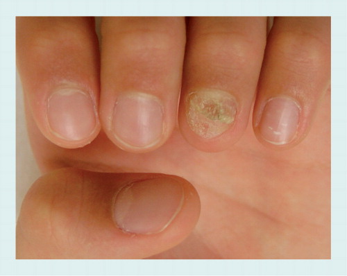
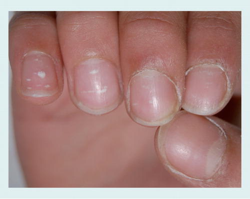
A single fingernail showing mild subungual hyperkeratosis and onycholysis, associated with periungual scaling. Note traumatic punctate leukonychia of the fifth fingernail.
Isolation of Candida indicates secondary colonization and not primary invasion.