Figures & data
(A) Clinical overview. (B) Close-up clinical image showing a pigmented lesion surrounded by a whitish halo. (C) With dermoscopy, the lesion appears almost completely involuted, with a homogeneous pattern, surrounded by a rim of scar-like depigmentation.
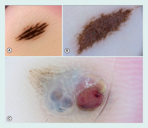
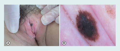
(A) A small acquired acral nevus showing a parallel furrow pattern. (B) Congenital acral nevus presenting a palpable area in the center of the lesion and displaying a grayish coloration by dermoscopy, corresponding to a dermal component of the nevus. At the periphery, a parallel furrow pattern can be detected. (C) Dermoscopy of an acral melanoma, arising on a pre-existing nevus. The melanoma was growing as a nodular component displaying a blue–white veil and an ulcerated red area.
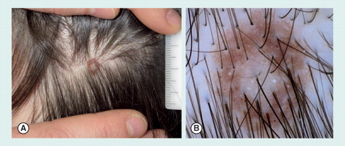
(A) Clinical view with presence of a white central area surrounded by a rim of brown pigmentation. (B) Under a dermoscope, the lesion shows a pigment network with perifollicular hypopigmentation, and scattered homogeneous brown globules. At the center, a hypopigmented, white area is detected.
(A) Clinical image showing a dark brown macule on the labia. (B) On dermoscopy, the lesion shows a mixed pattern with a homogeneous brown pigmentation and a central blue–grey area.
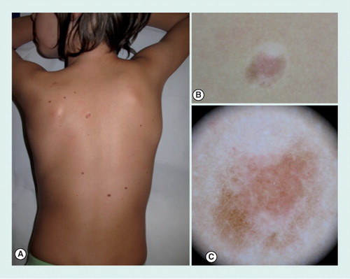
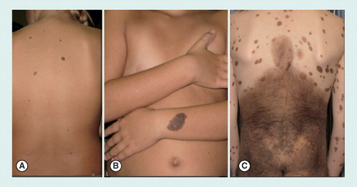
(A) Small (<1.5 cm diameter) congenital nevi on the back of an 8-year-old girl. (B) Medium size (1.5–20 cm) congenital nevus on the arm of an 8-year-old girl. (C) Giant congenital nevus (>20 cm), involving the area of the buttocks and the back, in a 9-year-old boy. Of note, the presence of multiple satellite nevi can be noticed.
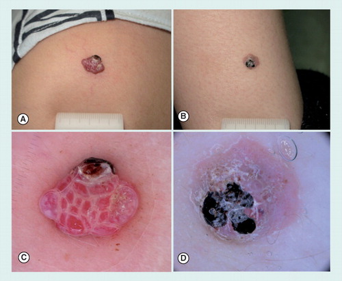
(A) A Spitz and (B) a Reed nevus arising, respectively, as a pink and black papule on the upper and lower arm of a 3-year-old boy. (C) Dermoscopy of nonpigmented Spitz nevus with dotted vessels and pinkish background. (D) Dermoscopy of Reed nevus, showing a globular pattern, composed by heavily pigmented brown-to-black globules.
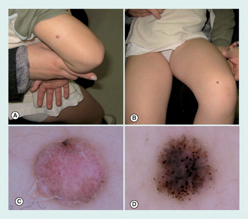
(A) A pyogenic granuloma and (B) an atypical Spitz tumor arising on the lower arm of two prepubertal children. Both lesions presented as pink to red nodules, partially covered by a blood crust. (C) Dermoscopy of pyogenic granuloma, displaying multiple red lacunes intersected by whitish lines. In the upper part of the lesion, a small ulcerated area is visible. (D) Aspecific dermoscopic pattern of an atypical Spitz tumor presenting a pink background with a few dotted vessels and a keratotic surface partially covered by a blood crust.