Figures & data
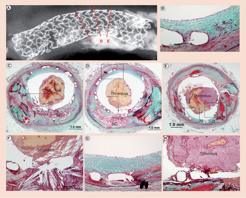
An 81-year-old male died suddenly 2 months after Taxus™ stent implantation in the left anterior descending artery for acute myocardial infaction. (A) Radiograph showing the area of plaque rupture (C, D & E) with three histologic sections. (B & D) Low-power sections of site of plaque rupture with (F) corresponding high powers. Note absence of fibrous cap and lack of stent coverage with overlay platelet-rich thrombus mixed with red cells. (D & E) Nonculprit sites with neointimal coverage of stent struts (boxed areas) and (G & B) persistant fibrin deposition. (E & H) Low and high powers of adjoining area of calcification at the site of plaque rupture (arrow). (H) Note fibrin coverage of stent struts, which are overlying an area of calcification with luminal platelet rich thrombus.
(B & E) High-power images of strut regions (H & E) showing presence of fibrin at 1 month and absence at 36 months. Note the strut outline is barely visible at 36 months. α actin-positive smooth muscle cells are observed in the neointima and media at (C) 1 month and (F) 36 months. (G) Complete degradation of the polymer strut with surrounding basophilic deposition of calcium (H & E). (H) Alcian blue positive proteoglycan infiltrated the matrix of the Bioabsorbable Vascular Solutions stent stent strut. (I) Calcification is seen around the degraded stent strut (von Kossa).
Modified and reproduced with permission from Citation[18].
![Figure 2. Representive histological sections of Bioabsorbable Vascular Solutions stent in pig coronary arteries removed at (A) 1 month and (D) 36 months (EVG staining).(B & E) High-power images of strut regions (H & E) showing presence of fibrin at 1 month and absence at 36 months. Note the strut outline is barely visible at 36 months. α actin-positive smooth muscle cells are observed in the neointima and media at (C) 1 month and (F) 36 months. (G) Complete degradation of the polymer strut with surrounding basophilic deposition of calcium (H & E). (H) Alcian blue positive proteoglycan infiltrated the matrix of the Bioabsorbable Vascular Solutions stent stent strut. (I) Calcification is seen around the degraded stent strut (von Kossa).Modified and reproduced with permission from Citation[18].](/cms/asset/b1913664-24e0-4625-b20e-88ff4c22a663/ierk_a_11210643_f0002_b.jpg)