Figures & data
Figure 1 Hypothetical age-size curves for fast-growing and slow-growing lichen genera, constructed at the beginning of the study (CitationBenedict, 1985: Fig. 44). Shaded boxes show radial growth rates. Triangles mark the transitions from sigmoidal to linear growth, predicted to occur when the radius of the thallus first equals the width of the peripheral growth zone. In the absence of a linear phase, growth will continue to follow the sigmoidal course indicated by the dashed lines.
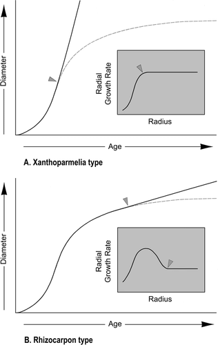
Figure 2 Map showing the locations of lichen-measurement sites. Route 72 is the Peak-to-Peak Scenic Byway.
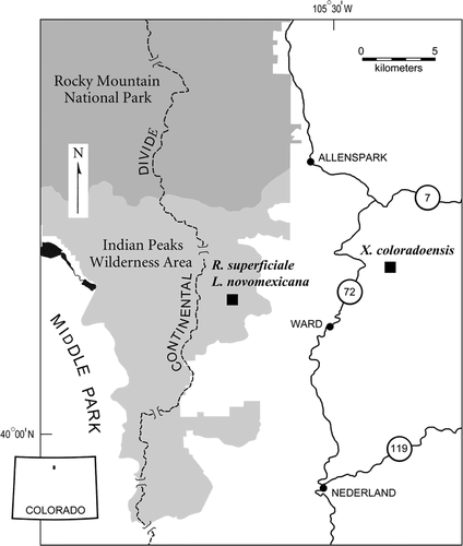
Figure 3 Catastrophic flood deposit on the south bank of South St. Vrain Creek, Boulder County, Colorado. Xanthoparmelia coloradoensis thalli were studied on unweathered outwash boulders near the crest of the flood bar. Snow fills a backwater slough at the base of the cliff from which the photograph was taken. Photo: 2 May 1994.
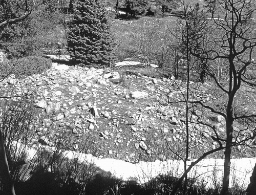
Table 1 Thallus characteristics, microenvironments, and growth rates.
Figure 4 Floor of the hanging valley on the southeast flank of Mount Audubon. Thalli of Rhizocarpon superficiale were measured on stable talus (A). Thalli of Lecanora novomexicana were measured in a depression behind the front of a lobate rock glacier (B). Areas of dark-colored debris are blown free of snow in winter and have mature lichen covers. Areas of light-colored debris experienced lichen snow-kill during the Late Holocene (CitationBenedict, 1999). Photo: 27 August 1990.
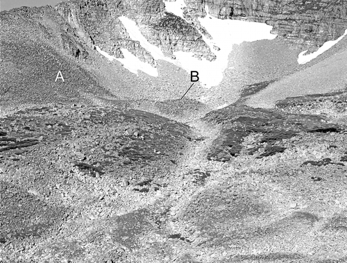
Figure 5 Photographs of an experimentally modified Xanthoparmelia coloradoensis thallus, showing reference crosses and aiming lines used to measure radial growth. By the conclusion of the two-year study, new lobes had begun to form along the knife-cut inner margin of the wedge. The lobes were widely spaced, however, and were too small to be clearly visible in the photograph.
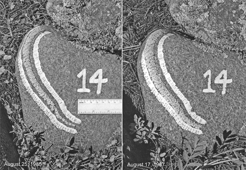
Figure 6 Radial growth of experimentally modified Xanthoparmelia coloradoensis thalli (1985–1987), measured to the tips of well-defined lobes. Black dots are 10-measurement averages except for the final dot, which is based on 13 measurements. The curves in – are fit by eye to the average values.
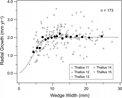
Figure 7 Radial growth of experimentally modified Lecanora novomexicana thalli (1985–1990), measured to the tips of marginal lobes. Black dots are 10-measurement averages except for the final dot, which is based on 11 measurements.
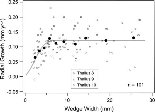
Figure 8 Radial growth of experimentally modified Rhizocarpon superficiale thalli (1986–2002), measured to the leading edges of marginal areolae and hypothalli. Black dots are 10-measurement averages except for the initial dot in each graph, which is the average of 8 or 5 measurements, depending upon sample size.
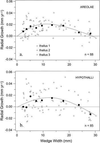
Figure 9 Thalli of Rhizocarpon superficiale photographed on 1 July 1985, before removing their interior portions. The central areas of both thalli were morphologically similar. But outer rings of areolate tissue that had not yet developed mature apothecia were strikingly different. Within these rings the areolae of fast-growing Thallus 2 were 2.6 times larger, and provided 26.3% more total photosynthetic area, than those of slow-growing Thallus 1 (). This allowed Thallus 2 to supply larger quantities of photosynthates to the growing hypothallus margin.
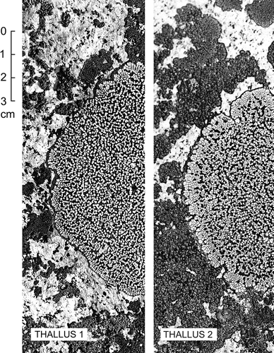
Table 2 Growth data for thalli used to construct .
Table 3 Comparison of areolae in slow-growing and fast-growing thalli.