Figures & data
FIGURE 1. Environmental scanning electron microscope (ESEM) images of pennate diatoms observed in QSD1, QSD2, and QSD3. (A) Pinnularia borealis, 59 × ∼12 µm. The inset shows nanoscale areolae within the striae, B) Hantzschia amphioxys, 58 × 9 µm, C) Pinnularia sp., 47 µm.
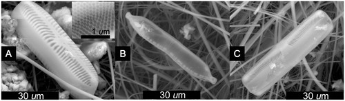
FIGURE 2. Aulacoseira valves (centric diatoms) observed in (A–C) QSD1, (D–F) QSD2, and (G–I) QSD3. Parts A, D, and F–I are Aulacoseira alpigena; others are Aulacoseira sp.
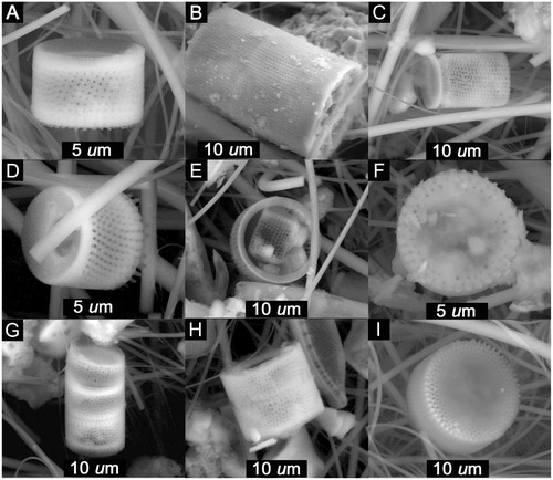
FIGURE 3. ESEM images of pennate diatoms found in QSD2. (A) Stauroneis sp. cf. S. phoenicenteron, 66 × 13.2 µm; (B) Stauroneis sp., 72 × 14 µm; (C) Caloneis sp., ∼66 × 10.8 µm; (D) Neidium bisulcatum, 46.2 × 7 µm; (E) Achnanthes sp. cf. A. taylorensis, 12.8 × 3.9 µm; (F) Pinnularia appendiculata, 33 × 5 µm.
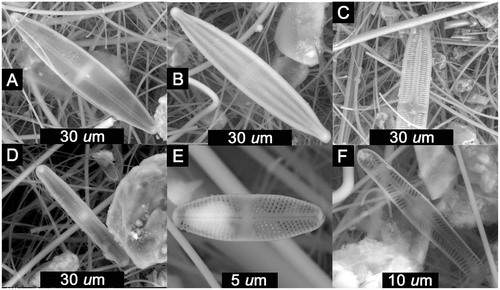
FIGURE 4. ESEM images of pennate diatoms found in QSD3. (A) Brachysira vitrea, 28 × 6.5 µm; (B) Placoneis elginensis, ∼30 × 10.3 µm; (C) Eunotia sp. cf. E. tenella, 19.7 × 6.3 µm; (D) Luticola sp. cf. L. nivalis, 31 × 10.5 µm; (E) Craticula sp., 65 × 15.5 µm.
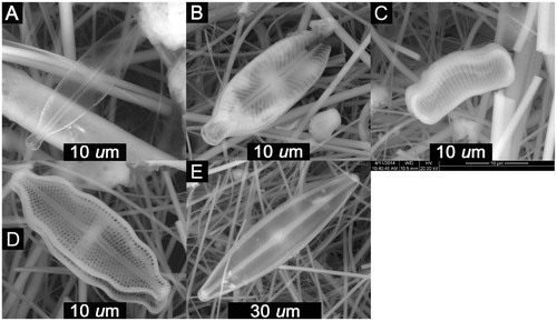
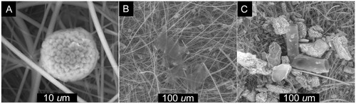
FIGURE 5. ESEM Images of (A) Volvox (green algae); (B) irregular e-beam transparent carbon masses; and (C) 60% carbon tubes.