Figures & data
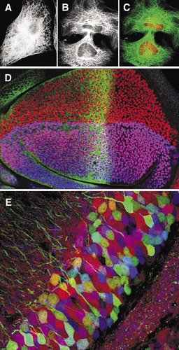
(A)–(C). Confocal imaging in the 1980s. (A) Wide-field epifluorescence image of a 3T3 cell immunofluorescently labeled with antitubulin. (B) Laser scanning confocal image of similar cell. (C) Double label confocal image of the same cell in (B); tubulin in green and nuclei labeled with propidium iodide in red. (D) Confocal imaging in the 1990s. Single optical sections collected simultaneously using a single krypton argon laser at three different excitation wavelengths—488 nm, 568 nm, and 647 nm—of a fruit fly third instar wing imaginal disk labeled for three genes involved with patterning the wing: cubitus interruptus (fluorescein, 496 nm) in green; vestigial (lissamine rhodamine, 572 nm) in red; and apterous (cyanine 5, 649 nm) in blue. (E) Confocal imaging in the 21st century. Brainbow image of mouse dentate gyrus (Citation21).