Figures & data
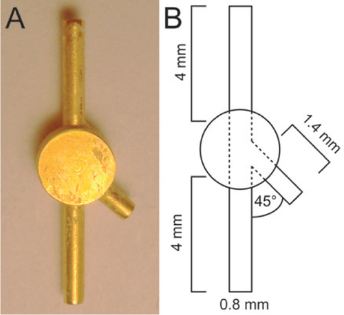
The Y-connection piece (Y-con) allows exchange of the pump reservoir of Alzet osmotic pumps by providing a pressure release exit. (A) Plain view of the Y-con. The Y-con consists of a central round plate and contains 3 adjusted channels. The two 4 mm long channels allow attachment of the new pump reservoir (bottom channel) and re-connection to the tubing that reaches the CNS cannula (top channel). The third 1.4 mm short channel (pressure release exit) enables equilibration of liquid pressure during surgery. (B) Scheme of the Y-con design, including length and angle details.
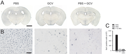
Y-con based pump reservoir exchange (first PBS, then GCV) and direct application of GCV lead to similar Iba1+ microglia ablation in the neocortex of TK mice. Three month old male TK mice received two weeks i.c.v. treatment of PBS, GCV, or PBS followed by two weeks GCV application (PBS + GCV) via pump exchange. (A) Direct i.c.v. infusion (left and middle panel) and pump reservoir replacement (right panel) using the Y-con, did not result in alterations of the lateral ventricle volume and revealed no tissue irritation. Calibration bar 2000 µm. (B) Iba1 immunohistochemistry in the neocortex of PBS-treated TK mice indicates a normal number of microglia cells (left panel), while direct application of GCV and treatment with PBS prior to GCV (PBS + GCV) via pump reservoir replacement resulted in almost complete microglia ablation (middle and right panel). Calibration bar: 100 µm. (C) Quantitative stereological analysis of Iba1+ cell number in the neocortex reveals a >90% ablation of microglia in TK mice after 2 weeks GCV treatment in comparison to the PBS treated control group (bars indicate the mean ± SEM; n = 3 mice/group). ANOVA indicates a significant difference between the three treatment groups (F(2,8) = 406.48; ***P < 0.001). Tukey HSD post-hoc analysis displays significant differences between the PBS and GCV treated TK mice.
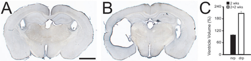
(A) PBS application (2 weeks) into the ventricle of 3 month old TK mice does not alter the size of the lateral ventricle system (n = 3). (B) Pump reservoir replacement applying direct reconnection of the tubing to the brain cannula (after initial 2 weeks PBS application) results in hydrocephalus in 3 month old TK mice 2 weeks after pump reservoir exchange (n = 3). Calibration bar: 1000 µm. (C) Quantitative stereological analysis of the ratio of lateral ventricle volume to total brain volume reveals a 2-fold increase (mean ± SEM; n = 3 mice/group); t-test indicates a significant difference between the groups (not replaced pump (nrp) and directly replaced pump (drp); t = 13.28, df = 4, ***P < 0.001).