Figures & data
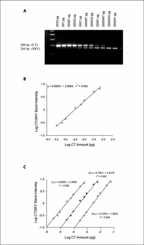
(A) Agarose gel electrophoresis of the coamplification of 0.1 ng of male genomic DNA (male DNA mixed with female DNA at a ratio of 1:104) with serial competitive template (CT) amounts ranging from 0.033 to 0.00001 pg. The gel shows two PCR products per lane, representing the CT (larger fragment, 300 bp) and SRY gene (smaller product, 266 bp). (B) Standard curve generated from the gel shown in panel A. The band intensity is assayed by scanning densitometry, and the graph is generated by plotting the log CT/SRY band intensity against the log CT amount. (C) Standard curves generated from amplifications containing three initial concentrations of male DNA substrate. Standard curve (i) represents the data from panel B (0.1 ng male DNA); (ii) and (iii) correspond to 1 and 10 ng male DNA, respectively.
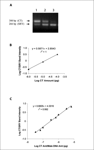
(A) Electrophoresis of 1000 ng of DNA extracted from a female liver post-transplantation submitted to standard PCR with SRY-specific primers and coamplified with competitive template (CT) of (Citation1) 0.0001, (Citation2) 0.00001, and (Citation3) 0.000001 pg, respectively. Scanning densitometry of the product bands generated ratios of 3.20, 0.83, and 0.21, respectively. (B) A conventional standard curve of the data shown in panel A, in which data are plotted as the log CT/SRY band intensity ratio versus the amount (amt) of CT used. (C) The master standard curve, generated from the conventional standard curve in panel B by the inclusion of male DNA amount added to the denominator on the x-axis.