

Figures & data
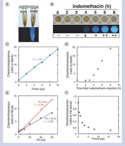
(A & B) We induced intestinal ulcer in mice injected with indomethacin, and harvested feces. (A) Feces in a microtube at 0 (con) and 6 h (indo) and (B) a portion of feces in a 96-well plate at different time points (0, 2, 3, 4, 5, 6 and 8 h) following the indomethacin injection. Fecal samples were mixed with a luminol solution and observed in dark field. The blue–white chemiluminescence became detectable at 4 h after the injection: -: negative; ±: dim; +: moderate; and ++: bright. (C) Different amounts of fecal samples from A were placed into a 96-well plate and mixed with 100 μl of a dilute luminol solution. Chemiluminescence intensities were measured by a microplate reader. We calculated the ratios of the chemiluminescence intensities of wells containing different amounts of feces to the chemiluminescence intensity of wells containing a luminol solution alone. Weights of feces containing occult blood positively correlated with the chemiluminescence ratios (r2 = 1.00). (D) We placed 5 μg of fecal samples from B, mixed with the luminol solution, and calculated the chemiluminescence ratios to the chemiluminescence intensity of wells containing feces from untreated mouse (time: 0 h). (E) We placed different amounts of hemoglobin (Hb) in 96-well plates in the absence (‘Hb alone’, open red circles) or the presence (‘Hb + feces’, closed purple circles) of 5 μg of control feces, followed by addition of the luminol solution. The chemiluminescence intensity of the luminol solution alone or with 5 μg of feces was used for the chemiluminescence ratios of ‘Hb alone’ or ‘Hb + feces’, respectively. Both Hb concentrations of ‘Hb alone’ or ‘Hb + feces’ samples positively correlated with the chemiluminescence ratios, although the ratios of the latter samples showed lower chemiluminescence ratios. (F) We placed different amounts of feces from normal mice (1–16 μg/well) into wells containing 50 ng of Hb, followed by addition of the luminol solution. We calculated the chemiluminescence ratios to the chemiluminescence intensity of wells containing 50 ng of Hb without feces. (A–F) We repeated each experiment at least three times.
Con: Control; Hb: Hemoglobin; Indo: Indomethacin.
Visual detection (A) and chemiluminescence measurement (B). (A) Visual detection: (1) Add a luminol dilute solution (dilute the original solution fivefold with deionized-distilled water [ddH2O]) to pooled feces in a microtube (left) or a piece of feces taken by a toothpick in a white 96-well plate (right); and (2) observe them in dark field. In the presence of blood, blue–white chemiluminescence is visible. (B) Chemiluminescence measurement: (1) Prepare fecal solutions with ddH2O (1 mg/ml) in a microtube; (2) vortex and centrifuge the tube for 2 min; (3) transfer 5 μl of the solution (containing 5 μg of feces) in a 96-well plate; (4) add 100 μl of a luminol dilute solution (diluted 500-fold with ddH2O) to each well; and (5) measure the chemiluminescence signal by a luminometer immediately. (C) Fecal blood scores based on the two luminol protocols. To calculate the chemiluminescence ratios, it is recommended to store the control fecal solution (1 mg/ml) derived from normal mice. Hb concentrations have been estimated by comparing with the results of feces mixed with standard Hb solutions.
ddH20: Deionized-distilled water; Hb: Hemoglobin.
![Figure 2. Schematic illustration of two protocols for detecting fecal occult blood by the luminol reaction.Visual detection (A) and chemiluminescence measurement (B). (A) Visual detection: (1) Add a luminol dilute solution (dilute the original solution fivefold with deionized-distilled water [ddH2O]) to pooled feces in a microtube (left) or a piece of feces taken by a toothpick in a white 96-well plate (right); and (2) observe them in dark field. In the presence of blood, blue–white chemiluminescence is visible. (B) Chemiluminescence measurement: (1) Prepare fecal solutions with ddH2O (1 mg/ml) in a microtube; (2) vortex and centrifuge the tube for 2 min; (3) transfer 5 μl of the solution (containing 5 μg of feces) in a 96-well plate; (4) add 100 μl of a luminol dilute solution (diluted 500-fold with ddH2O) to each well; and (5) measure the chemiluminescence signal by a luminometer immediately. (C) Fecal blood scores based on the two luminol protocols. To calculate the chemiluminescence ratios, it is recommended to store the control fecal solution (1 mg/ml) derived from normal mice. Hb concentrations have been estimated by comparing with the results of feces mixed with standard Hb solutions.ddH20: Deionized-distilled water; Hb: Hemoglobin.](/cms/asset/4e632788-c26f-4ea0-81b3-b81dc152ec23/ibtn_a_12360545_f0002.jpg)

