Figures & data
Table 1. Mononuclear cell recovery and viability for each donor.
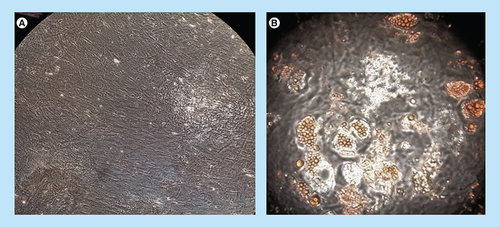
Example of one representative culture of mesenchymal stromal cells.
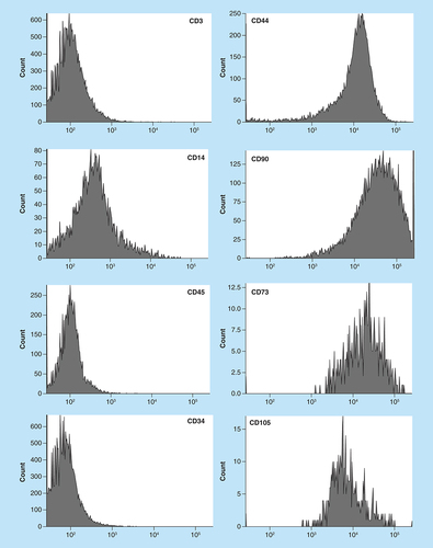
Table 2. Immunophenotype analysis of mesenchymal stromal cells obtained, for each surface antigen evaluated and standard deviation.
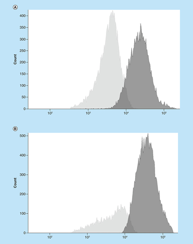
Proliferating responder cells are depicted in light gray, in the absence (A) and presence (B) of mesenchymal stromal cells. Dark gray represents nonproliferating cells.
(A) Negative control; (B) mesenchymal stromal cell-derived adypocytes with oil red O-stained droplets evident.