
Figures & data
Figure taken from Figure 1 from [Citation35] (original article is open access [CC-BY]).
CCD: Charged-coupled device.
![Figure 1. Helium–neon laser-induced thrombosis/thrombolysis (fibrinolysis) system.Figure taken from Figure 1 from [Citation35] (original article is open access [CC-BY]).CCD: Charged-coupled device.](/cms/asset/16c05042-e73e-4381-a303-f477b29f68ff/ifso_a_12364471_f0001.jpg)
(A) Thrombus size measurement of thrombus in mouse carotid artery. (B) Index of thrombogenicity measurement.
Figure taken from from [Citation35] (original article is open access [CC-BY]).
T: Thrombus; W: Vessel wall.
![Figure 2. Thrombosis in mouse carotid artery.(A) Thrombus size measurement of thrombus in mouse carotid artery. (B) Index of thrombogenicity measurement.Figure taken from Figure 1 from [Citation35] (original article is open access [CC-BY]).T: Thrombus; W: Vessel wall.](/cms/asset/de97a7c6-e2dc-417f-b0c2-542a3897c70d/ifso_a_12364471_f0002.jpg)
Thrombus area is calculated by delineating thrombus using computer (A). Subsequently, thrombus size (B) is obtained by multiplying gray scale and the area. Thrombolysis rate is compared with that at the start.
L: Lumen; T: Thrombus; V: Vessel wall.
Figure reproduced with permission from [Citation32] (Copyright © 2001, © 2001 S. Karger AG, Basel).
![Figure 3. Thrombolysis measurement in rat mesenteric microvessel.Thrombus area is calculated by delineating thrombus using computer (A). Subsequently, thrombus size (B) is obtained by multiplying gray scale and the area. Thrombolysis rate is compared with that at the start.L: Lumen; T: Thrombus; V: Vessel wall.Figure reproduced with permission from [Citation32] (Copyright © 2001, © 2001 S. Karger AG, Basel).](/cms/asset/33023e61-1ba1-418f-b7b1-0478431ddde9/ifso_a_12364471_f0003.jpg)
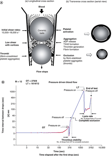
(A) Principle of ex vivo GTT. Platelets are activated under high shear condition at the upper gaps created along the inner of a conical plastic tube. Activated platelets form fibrin-stabilized platelet aggregates under low shear condition between two balls. Fibrin-stabilized platelet aggregates occlude the lower gaps. (B) Pattern obtained by GTT (GTT-3).
GTT: Global thrombosis test; LT: Lysis time OT: Occlusion time.
(A) Assessment of antithrombotic/prothrombotic activity by ex vivo test (GTT). (B) Assessment by in vivo test (He–Ne laser-induced thrombosis test). (A) Cabernet sauvignon. (B) Neo muscat.
(Figure taken from from [Citation50] (original article is open access [CC-BY]).
GTT: Global thrombosis test; LT: Lysis time OT: Occlusion time.
![Figure 5. Ex vivo and in vivo test.(A) Assessment of antithrombotic/prothrombotic activity by ex vivo test (GTT). (B) Assessment by in vivo test (He–Ne laser-induced thrombosis test). (A) Cabernet sauvignon. (B) Neo muscat.(Figure taken from Figure 1 from [Citation50] (original article is open access [CC-BY]).GTT: Global thrombosis test; LT: Lysis time OT: Occlusion time.](/cms/asset/a35a4658-6c11-4b33-ad29-ea34a6de47af/ifso_a_12364471_f0005.jpg)
Thrombotic and fibrinolytic activities in Westerners and Japanese, measured by GTT.
GTT: Global thrombosis test; J: Japanese; LT: Lysis time OT: Occlusion time; W: Westerners.
Reprinted from [Citation72] with permission from Elsevier.
![Figure 6. Comparison of thrombotic status between different races.Thrombotic and fibrinolytic activities in Westerners and Japanese, measured by GTT.GTT: Global thrombosis test; J: Japanese; LT: Lysis time OT: Occlusion time; W: Westerners.Reprinted from [Citation72] with permission from Elsevier.](/cms/asset/0cc8226d-d391-48b5-94c1-16b781b72469/ifso_a_12364471_f0006.jpg)
Enhanced thrombotic status in patients with cancers.
Figure taken from [Citation76]; was orally presented by Dr. Shioyama at The 25th Kinki Thrombosis Research Society Meeting. Osaka, Japan (2020).
![Figure 7. Thrombotic status of patients with and without cancers.Enhanced thrombotic status in patients with cancers.Figure taken from [Citation76]; was orally presented by Dr. Shioyama at The 25th Kinki Thrombosis Research Society Meeting. Osaka, Japan (2020).](/cms/asset/1e5c1fdb-cb7d-40c7-b372-d8728c2a725a/ifso_a_12364471_f0007.jpg)