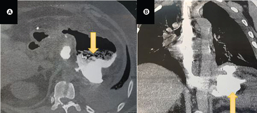
Figure 1. (A) CT scan axial view showing a left subphrenic collection (yellow arrow) communicating with the gastric sleeve.
(B) CT scan coronal view showing the left subphrenic collection (yellow arrow).
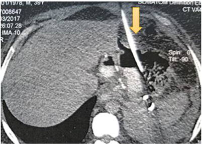
Figure 2. A coronal abdominal CT section showing the percutaneous radiological drainage (yellow arrow).
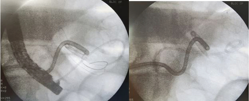
Figure 3. X-ray showing the endoscopic internal drainage with double pigtail plastic stent.
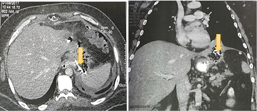
Figure 4. CT scan axial view showing a slight decrease in the size of the subphrenic collection with a persistent fistula and a spongiform formation with opaque serpiginous structures (yellow arrow) typical of a textiloma.
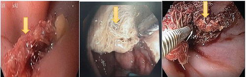
Figure 5. Upper endoscopy showing a narrowed cardia with a 10 mm loss of substance on the right edge blocked by a textiloma (yellow arrow).